This old version of Proteopedia is provided for student assignments while the new version is undergoing repairs. Content and edits done in this old version of Proteopedia after March 1, 2026 will eventually be lost when it is retired in about June of 2026.
Apply for new accounts at the new Proteopedia. Your logins will work in both the old and new versions.
Image:Pdx1.jpg
From Proteopedia
| Line 1: | Line 1: | ||
'''General structure of PDX-1 Homeodomain''' | '''General structure of PDX-1 Homeodomain''' | ||
| + | |||
| + | |||
This PDB structure corresponds to the PDX-1 homeodomain. This part of the transcription factor folds into three α-helices (pink color) and a flexible N-terminal arm (white-grey color). This homeodomain interacts with DNA (purple color). | This PDB structure corresponds to the PDX-1 homeodomain. This part of the transcription factor folds into three α-helices (pink color) and a flexible N-terminal arm (white-grey color). This homeodomain interacts with DNA (purple color). | ||
Current revision
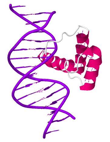
General structure of PDX-1 Homeodomain
This PDB structure corresponds to the PDX-1 homeodomain. This part of the transcription factor folds into three α-helices (pink color) and a flexible N-terminal arm (white-grey color). This homeodomain interacts with DNA (purple color).
References : http://www.rcsb.org/pdb/explore/jmol.do?structureId=2H1K&bionumber=1
File history
Click on a date/time to view the file as it appeared at that time.
| Date/Time | User | Dimensions | File size | Comment | |
|---|---|---|---|---|---|
| (current) | 10:11, 31 December 2013 | Megane Denu (Talk | contribs) | 363×466 | 24 KB |
- Edit this file using an external application
See the setup instructions for more information.
Links
The following pages link to this file:
