This old version of Proteopedia is provided for student assignments while the new version is undergoing repairs. Content and edits done in this old version of Proteopedia after March 1, 2026 will eventually be lost when it is retired in about June of 2026.
Apply for new accounts at the new Proteopedia. Your logins will work in both the old and new versions.
Introduction to protein structure
From Proteopedia
(Difference between revisions)
| Line 20: | Line 20: | ||
== Ways of representing protein structure == | == Ways of representing protein structure == | ||
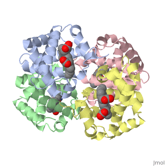
| - | Protein structures can be displayed in many different ways. In <scene name='57/575866/Spacefill_segment/1'>spacefilling</scene> models, all of the non-hydrogen atoms are shown as spheres with their van der Waals radii. In the <scene name='57/575866/Ball_and_stick_segment/1'>ball and stick</scene> model, the atoms are shown as smaller balls, connected by sticks; this is further simplified in the <scene name='57/575866/Stick_segment/1'>stick</scene> model, which only shows the bonds between atoms. <scene name='57/575866/Backbone/2'>Backbone</scene> shows only the N-Calpha-C=O repeating unit; the <scene name='57/575866/Cartoon/ | + | Protein structures can be displayed in many different ways. In <scene name='57/575866/Spacefill_segment/1'>spacefilling</scene> models, all of the non-hydrogen atoms are shown as spheres with their van der Waals radii. In the <scene name='57/575866/Ball_and_stick_segment/1'>ball and stick</scene> model, the atoms are shown as smaller balls, connected by sticks; this is further simplified in the <scene name='57/575866/Stick_segment/1'>stick</scene> model, which only shows the bonds between atoms. <scene name='57/575866/Backbone/2'>Backbone</scene> shows only the N-Calpha-C=O repeating unit; the <scene name='57/575866/Cartoon/4'>cartoon</scene> representation shows the secondary structures. |
'''Questions based upon these scenes:''' | '''Questions based upon these scenes:''' | ||
| Line 40: | Line 40: | ||
Clicking the right mouse button brings up an extensive menu. This exercise will use commands in the style, color,zoom, measurements, and set picking categories. | Clicking the right mouse button brings up an extensive menu. This exercise will use commands in the style, color,zoom, measurements, and set picking categories. | ||
| - | We will begin with the <scene name='57/575866/Cartoon/ | + | We will begin with the <scene name='57/575866/Cartoon/4'>cartoon</scene> structure of an alpha helix from hemoglobin. From this view, can you determine: |
--The number of amino acids per turn? | --The number of amino acids per turn? | ||
| Line 49: | Line 49: | ||
Rotate the helix so that you are looking down the helix. What does the middle of the helix look like? | Rotate the helix so that you are looking down the helix. What does the middle of the helix look like? | ||
| - | Right click on the mouse, choose | + | Right click on the mouse, choose style, then scheme, then CPK spacefill. |
| + | |||
| + | What does the middle of the helix look like? | ||
| + | Which view is more representative of the true structure of the molecule? | ||
| + | |||
| + | Let's try changing to another view. Right click on the mouse, choose style, then scheme, then ball and stick. Based upon what you know about peptide composition or by holding the mouse over the atoms determine the color scheme: | ||
| + | red = | ||
| + | |||
| + | black = | ||
| + | |||
| + | blue = | ||
| + | |||
| + | Notice that hydrogens are not shown on this model. Xray crystallography is not able to resolve hydrogens, so they are omitted from the images. This also simplifies the data set, as there are many fewer atoms to position. | ||
| + | |||
| + | |||
| + | |||
| + | |||
-- | -- | ||
</StructureSection> | </StructureSection> | ||
Revision as of 13:58, 16 January 2014
Levels of Protein Structure
| |||||||||||
Proteopedia Page Contributors and Editors (what is this?)
Ann Taylor, Joel L. Sussman, Alexander Berchansky, Eric Martz, Israel Hanukoglu, Jaime Prilusky, Nick Kenworthy
