Sandbox Reserved 932
From Proteopedia
m |
|||
| Line 21: | Line 21: | ||
| - | Three-finger toxins (3FTXs) are the most common family of snake venom proteins; these venoms can be found in elapid, colubrid and hydrophiid snakes and include toxins such as α-cobratoxin and α-bungarotoxin. 3FTXs are non-enzymatic proteins which form a structurally conserved superfamily whose members all share a highly conserved structure. <ref name=Dufton></ref><ref name=Endo></ref>The core structure of 3FTXs is formed by three <scene name='57/579702/Three_fingers/1'>three β-stranded polypeptide loops</scene> joined together by four conserved disulphide bridges located in the <scene name='57/579702/3ftx_beta_strands/2'>core</scene> of the protein. Despite the similarities in the structure of different toxins belonging to the family, the 3FTXs from various venoms have a variety of receptors/acceptors and exhibit differential responses in their targets. The members of the family can vary slightly in: the length and type of twists of the tree loops; the length and type of turns of the N and C-terminal tail and the amount of β-sheets in the overall structure. These differences allow for the specificity and toxicity of the proteins to their targets. <ref name=Pawlak/> | + | Three-finger toxins (3FTXs) are the most common family of snake venom proteins; these venoms can be found in elapid, colubrid and hydrophiid snakes and include toxins such as α-cobratoxin and α-bungarotoxin. 3FTXs are non-enzymatic proteins which form a structurally conserved superfamily whose members all share a highly conserved structure. <ref name=Dufton></ref><ref name=Endo></ref>The core structure of 3FTXs is formed by three <scene name='57/579702/Three_fingers/1'>three β-stranded polypeptide loops</scene> joined together by four conserved disulphide bridges located in the <scene name='57/579702/3ftx_beta_strands/2'>core</scene> of the protein. Despite the similarities in the structure of different toxins belonging to the family, the 3FTXs from various venoms have a variety of receptors/acceptors and exhibit differential responses in their targets. The members of the family can vary slightly in: the length and type of twists of the tree loops; the length and type of turns of the N and C-terminal tail and the amount of β-sheets in the overall structure. These differences allow for the specificity and toxicity of the proteins to their targets. <ref name=Pawlak></ref> |
Denmotoxin shares approximately 30% sequence similarity with other 3FTXs with an exception of exhibiting approximately 50% sequence similarity with another colubrid snake venom α-colubritoxin. Despite the relatively low sequence similarity, denmotoxin possesses all the residues needed to maintain the 3 finger fold. A large part of the sequence similarity between denmotoxin and other 3FTXs is due to the highly conserved disulphides and a number of structurally important residues. <ref name=Pawlak/> | Denmotoxin shares approximately 30% sequence similarity with other 3FTXs with an exception of exhibiting approximately 50% sequence similarity with another colubrid snake venom α-colubritoxin. Despite the relatively low sequence similarity, denmotoxin possesses all the residues needed to maintain the 3 finger fold. A large part of the sequence similarity between denmotoxin and other 3FTXs is due to the highly conserved disulphides and a number of structurally important residues. <ref name=Pawlak/> | ||
| Line 45: | Line 45: | ||
Biochemistry of denmotoxin is unique for its taxon specificity to bird nicotinic acetylcholine receptors (nAChR). Binding of denmotoxin to chick muscle AChR (α1βγδ) is a highly irreversible whereas interaction with identical subunit assembly in mouse AChR is reversible. The reversible binding allows the receptor to function properly, but in the case of irreversible binding nAChR is prevented of natural agonist activation. Previous studies <ref>Samson. A. O. & Levitt M., [http://pubs.acs.org/doi/abs/10.1021/bi702272j "Inhibition Mechanism of the Acetylcholine Receptor by α-Neurotoxins as Revealed by Normal-Mode Dynamics"], ''Biochemistry, 2008, 47 (13), pp 4065–4070'', March 8, 2008. Retrieved May 19, 2014.</ref> with 3FTXs have shown that the binding of toxin leads to “locking down” of the nACh receptor, preventing required conformational change for ion channel activation and induction of signal. <ref name=Pawlak/> | Biochemistry of denmotoxin is unique for its taxon specificity to bird nicotinic acetylcholine receptors (nAChR). Binding of denmotoxin to chick muscle AChR (α1βγδ) is a highly irreversible whereas interaction with identical subunit assembly in mouse AChR is reversible. The reversible binding allows the receptor to function properly, but in the case of irreversible binding nAChR is prevented of natural agonist activation. Previous studies <ref>Samson. A. O. & Levitt M., [http://pubs.acs.org/doi/abs/10.1021/bi702272j "Inhibition Mechanism of the Acetylcholine Receptor by α-Neurotoxins as Revealed by Normal-Mode Dynamics"], ''Biochemistry, 2008, 47 (13), pp 4065–4070'', March 8, 2008. Retrieved May 19, 2014.</ref> with 3FTXs have shown that the binding of toxin leads to “locking down” of the nACh receptor, preventing required conformational change for ion channel activation and induction of signal. <ref name=Pawlak/> | ||
| - | There are no significant differences in the sequence of functionally important loops A-F of nAChR in mice and chicks. However in the prior region of loop F, chicks have several changes in their amino acid composition leading to introduction of positive charge spatially in the front of the functionally active loop F. This might have important functionality in the attraction and binding of denmotoxin specifically to bird nAChRs. However, no crystal structure of denmotoxin binding to nAChR has been resolved; thus denmotoxin could also utilise a yet uncharacterised binding site in the receptor | + | There are no significant differences in the sequence of functionally important loops A-F of nAChR in mice and chicks. However in the prior region of loop F, chicks have several changes in their amino acid composition leading to introduction of positive charge spatially in the front of the functionally active loop F. This might have important functionality in the attraction and binding of denmotoxin specifically to bird nAChRs. However, no crystal structure of denmotoxin binding to nAChR has been resolved; thus denmotoxin could also utilise a yet uncharacterised binding site in the receptor. <ref name=Pawlak/> |
Revision as of 15:01, 18 May 2014
| This Sandbox is Reserved from 01/04/2014, through 30/06/2014 for use in the course "510042. Protein structure, function and folding" taught by Prof Adrian Goldman, Tommi Kajander, Taru Meri, Konstantin Kogan and Juho Kellosalo at the University of Helsinki. This reservation includes Sandbox Reserved 923 through Sandbox Reserved 947. |
To get started:
More help: Help:Editing |
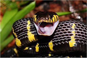
B. Dendrophila monomeric toxin (Denmotoxin) is the primary protein of snake venom used by Boiga dendrophila (fig. 1). This colubrid snake lives in Southest Asian lowland rainforest and mangrove swamps using birds as its primary prey. It belongs to one of the most well characterized snake venom protein families is the Three-finger-toxins (3FTX). These proteins consist of three β-stranded finger-like polypeptide loops stabilized by four disulphide bridges on the surface of a globular core. In non-convential 3TFXs a fifth disulphide bridge can be present as is the case in Denmotoxin. The crystal structure of denmotoxin was solved to 1.9Å by molecular replacement method. [1][2]
Denmotoxin binds specifically to bird muscle nicotinic acetylcholine receptors preventing their normal function in signal transduction. This taxon specifity is reached by unique structural differences to other 3FTXs such as changes in the suggested binding loop of the protein. [3]
Denmotoxin
| |||||||||||
Additional Information
Authors: Karoliina Hassi & Tommi Kotila, University of Helsinki
References
- ↑ Dufton M.J. & Hider R.C., "Structure and pharmacology of elapid cytotoxins", Pharmacol Ther. 1988;36(1):1-40, 1988. Retrieved May 19, 2014.
- ↑ Endo T. & Tamiya N., Structure-function relationship of postsynaptic neurotoxins from snake venoms, Snake Toxins, pp. 165-222., 1991. Retrieved May 19, 2014.
- ↑ 3.0 3.1 3.2 3.3 3.4 3.5 3.6 3.7 Pawlak J. et al., "Denmotoxin, a Three-finger Toxin from the Colubrid Snake Boiga dendrophila (Mangrove Catsnake) with Bird-specific Activity", The Journal of Biological Chemistry: 281: 29030-29041, September 29, 2006. Retrieved May 19, 2014.
- ↑ Huang S. et al., "Complex between α-bungarotoxin and an α7 nicotinic receptor ligand-binding domain chimaera", Biochem J. 454(2): 303–310., Septemper 1, 2013. Retrieved May 19, 2014.
- ↑ Samson. A. O. & Levitt M., "Inhibition Mechanism of the Acetylcholine Receptor by α-Neurotoxins as Revealed by Normal-Mode Dynamics", Biochemistry, 2008, 47 (13), pp 4065–4070, March 8, 2008. Retrieved May 19, 2014.

![Figure 2. Three fingers formed by three peptide loops (F1, F2 & F3) [PDB=2H5F]](/wiki/images/thumb/3/3f/Kolme_sormea_figure_text.png/300px-Kolme_sormea_figure_text.png)
![Figure 3. Suggested interaction of α-bungarotoxin in binding pocket of nAChR (simplified figure) [PDB=4HQP]](/wiki/images/thumb/d/d7/Bungarotoxin_interaction_with_nACHr_figure_text.png/300px-Bungarotoxin_interaction_with_nACHr_figure_text.png)
