1iat
From Proteopedia
| Line 4: | Line 4: | ||
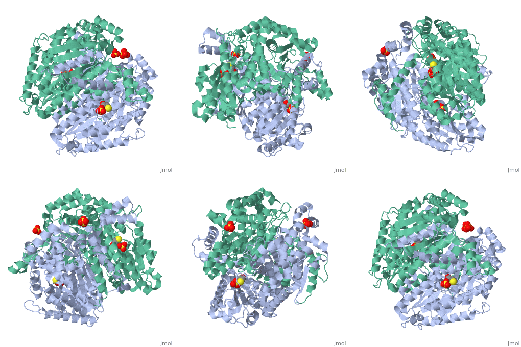
|PDB= 1iat |SIZE=350|CAPTION= <scene name='initialview01'>1iat</scene>, resolution 1.62Å | |PDB= 1iat |SIZE=350|CAPTION= <scene name='initialview01'>1iat</scene>, resolution 1.62Å | ||
|SITE= | |SITE= | ||
| - | |LIGAND= <scene name='pdbligand= | + | |LIGAND= <scene name='pdbligand=BME:BETA-MERCAPTOETHANOL'>BME</scene>, <scene name='pdbligand=SO4:SULFATE+ION'>SO4</scene> |
| - | |ACTIVITY= [http://en.wikipedia.org/wiki/Glucose-6-phosphate_isomerase Glucose-6-phosphate isomerase], with EC number [http://www.brenda-enzymes.info/php/result_flat.php4?ecno=5.3.1.9 5.3.1.9] | + | |ACTIVITY= <span class='plainlinks'>[http://en.wikipedia.org/wiki/Glucose-6-phosphate_isomerase Glucose-6-phosphate isomerase], with EC number [http://www.brenda-enzymes.info/php/result_flat.php4?ecno=5.3.1.9 5.3.1.9] </span> |
|GENE= GPI ([http://www.ncbi.nlm.nih.gov/Taxonomy/Browser/wwwtax.cgi?mode=Info&srchmode=5&id=9606 Homo sapiens]) | |GENE= GPI ([http://www.ncbi.nlm.nih.gov/Taxonomy/Browser/wwwtax.cgi?mode=Info&srchmode=5&id=9606 Homo sapiens]) | ||
| + | |DOMAIN= | ||
| + | |RELATEDENTRY=[[1dqr|1DQR]] | ||
| + | |RESOURCES=<span class='plainlinks'>[http://oca.weizmann.ac.il/oca-docs/fgij/fg.htm?mol=1iat FirstGlance], [http://oca.weizmann.ac.il/oca-bin/ocaids?id=1iat OCA], [http://www.ebi.ac.uk/pdbsum/1iat PDBsum], [http://www.rcsb.org/pdb/explore.do?structureId=1iat RCSB]</span> | ||
}} | }} | ||
| Line 16: | Line 19: | ||
==Disease== | ==Disease== | ||
| - | Known | + | Known disease associated with this structure: Hemolytic anemia due to glucosephosphate isomerase deficiency OMIM:[[http://www.ncbi.nlm.nih.gov/entrez/dispomim.cgi?id=172400 172400]], Hydrops fetalis, one form OMIM:[[http://www.ncbi.nlm.nih.gov/entrez/dispomim.cgi?id=172400 172400]] |
==About this Structure== | ==About this Structure== | ||
| Line 32: | Line 35: | ||
[[Category: Pearce, J.]] | [[Category: Pearce, J.]] | ||
[[Category: Read, J A.]] | [[Category: Read, J A.]] | ||
| - | [[Category: BME]] | ||
| - | [[Category: SO4]] | ||
[[Category: glycolysis enzyme/neurotrophic growth factor/cytokine]] | [[Category: glycolysis enzyme/neurotrophic growth factor/cytokine]] | ||
[[Category: isomerase]] | [[Category: isomerase]] | ||
[[Category: two alpha/beta domain]] | [[Category: two alpha/beta domain]] | ||
| - | ''Page seeded by [http://oca.weizmann.ac.il/oca OCA ] on | + | ''Page seeded by [http://oca.weizmann.ac.il/oca OCA ] on Sun Mar 30 21:17:07 2008'' |
Revision as of 18:17, 30 March 2008
| |||||||
| , resolution 1.62Å | |||||||
|---|---|---|---|---|---|---|---|
| Ligands: | , | ||||||
| Gene: | GPI (Homo sapiens) | ||||||
| Activity: | Glucose-6-phosphate isomerase, with EC number 5.3.1.9 | ||||||
| Related: | 1DQR
| ||||||
| Resources: | FirstGlance, OCA, PDBsum, RCSB | ||||||
| Coordinates: | save as pdb, mmCIF, xml | ||||||
CRYSTAL STRUCTURE OF HUMAN PHOSPHOGLUCOSE ISOMERASE/NEUROLEUKIN/AUTOCRINE MOTILITY FACTOR/MATURATION FACTOR
Contents |
Overview
Phosphoglucose isomerase (PGI) is a multifunctional protein, which, inside the cell, functions as a housekeeping enzyme of glycolysis and gluconeogenesis and, outside the cell, exerts wholly unrelated cytokine properties. We have determined the structure of human PGI to a resolution of 1.6 A using X-ray crystallography. The structure is highly similar to other PGIs, especially the architecture of the active site. Fortuitous binding of a sulphate molecule from the crystallisation solution has facilitated an accurate description of the substrate phosphate-binding site. Comparison with both native and inhibitor-bound rabbit PGI structures shows that two loops move closer to the active site upon binding inhibitor. Interestingly, the human structure most closely resembles the inhibitor-bound structure, suggesting that binding of the phosphate moiety of the substrate may trigger this conformational change. We suggest a new mechanism for catalysis that uses Glu357 as the base catalyst for the isomerase reaction rather than His388 as proposed previously. The human PGI structure has also provided a detailed framework with which to map mutations associated with non-spherocytic haemolytic anaemia.
Disease
Known disease associated with this structure: Hemolytic anemia due to glucosephosphate isomerase deficiency OMIM:[172400], Hydrops fetalis, one form OMIM:[172400]
About this Structure
1IAT is a Single protein structure of sequence from Homo sapiens. Full crystallographic information is available from OCA.
Reference
The crystal structure of human phosphoglucose isomerase at 1.6 A resolution: implications for catalytic mechanism, cytokine activity and haemolytic anaemia., Read J, Pearce J, Li X, Muirhead H, Chirgwin J, Davies C, J Mol Biol. 2001 Jun 1;309(2):447-63. PMID:11371164
Page seeded by OCA on Sun Mar 30 21:17:07 2008

