Monoclonal Antibody
From Proteopedia
(Difference between revisions)
| Line 2: | Line 2: | ||
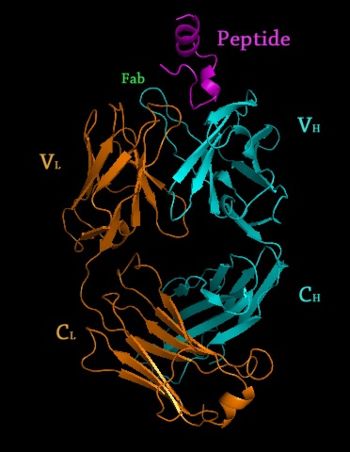
[[Image: Rituxan2.jpg|350px|left|thumb| Crystal Structure of Rituximab Fab in complex with an epitope peptide [[2osl]]]] | [[Image: Rituxan2.jpg|350px|left|thumb| Crystal Structure of Rituximab Fab in complex with an epitope peptide [[2osl]]]] | ||
{{Clear}} | {{Clear}} | ||
| + | |||
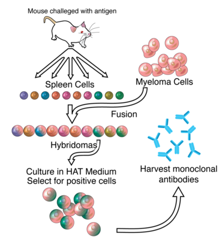
| + | '''Monoclonal antibodies''' are [[immunoglobulins]] produced by a single clone of cells and therefore a single pure homogeneous type of antibody. For structural information on immunoglobulins, see: [[Antibody]] The development of antibodies has revolutionized much of the science world both in the lab and in the medical world in treating cancer and autoimmune disorders. Humanized mouse antibody (hmFab) is a modified mFab which resembles more hFab. Catalytic antibody is a monoclonal antibody with catalytic activity. | ||
| + | |||
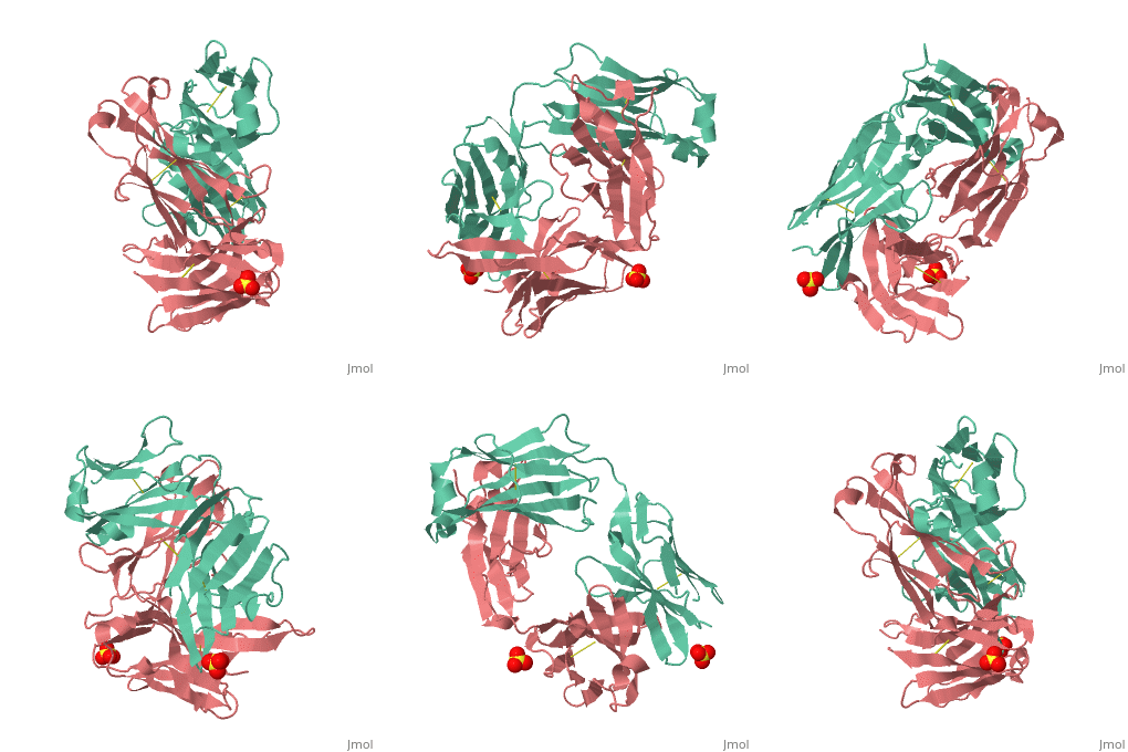
<scene name='Monoclonal_Antibody/Opening_monoclonal_antibody/2'>Crystal Structure of the Fab Fragment of Matuzumab</scene>, [[3c08]]. Light (blue) and heavy (yellow) chains. | <scene name='Monoclonal_Antibody/Opening_monoclonal_antibody/2'>Crystal Structure of the Fab Fragment of Matuzumab</scene>, [[3c08]]. Light (blue) and heavy (yellow) chains. | ||
| - | '''Monoclonal antibodies''' are [[immunoglobulins]] produced by a single clone of cells and therefore a single pure homogeneous type of antibody. For structural information on immunoglobulins, see: [[Antibody]] The development of antibodies has revolutionized much of the science world both in the lab and in the medical world in treating cancer and autoimmune disorders. Humanized mouse antibody (hmFab) is a modified mFab which resembles more hFab. Catalytic antibody is a monoclonal antibody with catalytic activity. | ||
<br /> | <br /> | ||
| Line 19: | Line 21: | ||
==Newer Monoclonal Technology== | ==Newer Monoclonal Technology== | ||
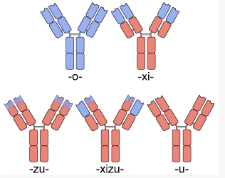
Early forms of monoclonal antibodies (murine antibodies) were problematic for therapeutic use because they were mouse antibodies, not human antibodies. When injected into humans, the antibodies would either be rapidly cleared from the body or worse, result in systemic inflammatory effects that were harmful. The human immune system recognized these mouse antibodies as foreign pathogens and rapidly produced [http://en.wikipedia.org/wiki/HAMA human anti-mouse antibodies] and unleashed the immune system on these invading pathogens. <ref>PMID:10835540</ref> To avoid this immune response, antibodies that the body recognized as native had to be created. Murine antibodies typically have a “mo” before the “mab” in their name, as is the case with Tositumomab (Marketed as Bexxar by GlaxoSmithKline), a drug used to treat lymphoma. | Early forms of monoclonal antibodies (murine antibodies) were problematic for therapeutic use because they were mouse antibodies, not human antibodies. When injected into humans, the antibodies would either be rapidly cleared from the body or worse, result in systemic inflammatory effects that were harmful. The human immune system recognized these mouse antibodies as foreign pathogens and rapidly produced [http://en.wikipedia.org/wiki/HAMA human anti-mouse antibodies] and unleashed the immune system on these invading pathogens. <ref>PMID:10835540</ref> To avoid this immune response, antibodies that the body recognized as native had to be created. Murine antibodies typically have a “mo” before the “mab” in their name, as is the case with Tositumomab (Marketed as Bexxar by GlaxoSmithKline), a drug used to treat lymphoma. | ||
| - | |||
| - | <scene name='Antibody/Rituxan_starting_scene/1'>Crystal structure of the Rituximab Fab in complex with an epitope peptide</scene> ([[2osl]]). | ||
===Chimeric Antibodies=== | ===Chimeric Antibodies=== | ||
| + | <scene name='Antibody/Rituxan_starting_scene/1'>Crystal structure of the Rituximab Fab in complex with an epitope peptide</scene> ([[2osl]]). | ||
| + | |||
[[Image: Chimera.PNG |350px|left|thumb| Different Types of Antibodies]] | [[Image: Chimera.PNG |350px|left|thumb| Different Types of Antibodies]] | ||
{{Clear}} | {{Clear}} | ||
| Line 32: | Line 34: | ||
The major drawback with chimeric antibody production is it still involves using rodents as the original source of antigen binding, and requires significant effort to identify, purify and create the fusion antibody. Applying a technique called [http://en.wikipedia.org/wiki/Phage_display phage display] to monoclonal antibody production expedites this entire process. In phage display, DNA coding the binding region of the antibody is ligated into the genes of a phage which code for proteins that are expressed on the protein coat of the virus. This phage gene hybrid is then transformed into bacterial cells. The virus hijacks the bacterial replication machinery, producing vast quantities of phage. By using many DNA fragments that are slightly different, a library of slightly different antibodies can be expressed on the outside of these phages, one type of antibody per phage. These antibody covered phages can then be selected for by running them through a column with the protein target immobilized on the column. Once the phages that bind the target protein well are identified, they can be used to reinfect bacterial cells and to isolate the DNA of antibodies that code for those antibodies with the greatest affinity for the target. Once the best antibodies are identified, they can be combined with human antibody scaffolds or human Fc regions as above to create humanized antibodies. <ref>pmid:1896445</ref> <ref>PMID:4001944</ref>Antibodies created through phage display have a “u” before the “mab” in their name to indicate their chain origins as is the case for [http://en.wikipedia.org/wiki/Adalimumab Adalimumab] ([http://www.humira.com/ HUMIRA]) | The major drawback with chimeric antibody production is it still involves using rodents as the original source of antigen binding, and requires significant effort to identify, purify and create the fusion antibody. Applying a technique called [http://en.wikipedia.org/wiki/Phage_display phage display] to monoclonal antibody production expedites this entire process. In phage display, DNA coding the binding region of the antibody is ligated into the genes of a phage which code for proteins that are expressed on the protein coat of the virus. This phage gene hybrid is then transformed into bacterial cells. The virus hijacks the bacterial replication machinery, producing vast quantities of phage. By using many DNA fragments that are slightly different, a library of slightly different antibodies can be expressed on the outside of these phages, one type of antibody per phage. These antibody covered phages can then be selected for by running them through a column with the protein target immobilized on the column. Once the phages that bind the target protein well are identified, they can be used to reinfect bacterial cells and to isolate the DNA of antibodies that code for those antibodies with the greatest affinity for the target. Once the best antibodies are identified, they can be combined with human antibody scaffolds or human Fc regions as above to create humanized antibodies. <ref>pmid:1896445</ref> <ref>PMID:4001944</ref>Antibodies created through phage display have a “u” before the “mab” in their name to indicate their chain origins as is the case for [http://en.wikipedia.org/wiki/Adalimumab Adalimumab] ([http://www.humira.com/ HUMIRA]) | ||
| + | ==Glycosylation of Monoclonal Antibodies== | ||
<scene name='Monoclonal_Antibody/Monoclonal_antibody_open/1'>Fc Fragment of Rituximab</scene> bound to a Protein Called Z34C,([[1l6x]]). | <scene name='Monoclonal_Antibody/Monoclonal_antibody_open/1'>Fc Fragment of Rituximab</scene> bound to a Protein Called Z34C,([[1l6x]]). | ||
| - | ==Glycosylation of Monoclonal Antibodies== | ||
Once an antibody gene is designed to contain the ideal sequence, the antibody then must be mass produced. An ideal system of production would be in bacterial cells because of the rapidity with which they replicate and ease of use. Unfortunately, bacterial cells do not glycosylate proteins the same way mammalian cells do. Since <scene name='Monoclonal_Antibody/Monoclonal_antibody_glycosylat/2'>protein glycosylation </scene>is critical for the immune system to recognize a protein as native, thus allowing the protein to stay in circulation in the human body without creating an immune response, antibody production had to be completed in cells that glycosylate the antibody appropriately. Chinese Hamster Ovary (CHO) cells are one type of cell that is able to glycosylate antibodies appropriately, but they are difficult to work with and relatively expensive at an industrial scale. Regardless, they are the most commonly used mammalian hosts for industrial production of recombinant protein therapeutics. <ref>PMID:15529164</ref> More recently, work by Dr. Tillman Gerngross, et al. have demonstrated that yeast can be genetically manipulated to produce human glycosylation on expressed antibodies. This is an important discovery because yeast replicate much faster than CHO cells, making them more ideal as hosts for industrial scale production of therapeutic proteins. <ref>PMID:12702754</ref> Gerngross’s startup company [http://www.glycofi.com GlycoFi], which was based around this platform was [http://www.glycofi.com/news/050906.html purchased by Merck in 2006] and the technology is being employed in Merck’s pipeline. Gerngross’s new company, [www.adimab.com Adimab], also uses similar technology to produce human glycosylated proteins for the pharmaceutical industry. | Once an antibody gene is designed to contain the ideal sequence, the antibody then must be mass produced. An ideal system of production would be in bacterial cells because of the rapidity with which they replicate and ease of use. Unfortunately, bacterial cells do not glycosylate proteins the same way mammalian cells do. Since <scene name='Monoclonal_Antibody/Monoclonal_antibody_glycosylat/2'>protein glycosylation </scene>is critical for the immune system to recognize a protein as native, thus allowing the protein to stay in circulation in the human body without creating an immune response, antibody production had to be completed in cells that glycosylate the antibody appropriately. Chinese Hamster Ovary (CHO) cells are one type of cell that is able to glycosylate antibodies appropriately, but they are difficult to work with and relatively expensive at an industrial scale. Regardless, they are the most commonly used mammalian hosts for industrial production of recombinant protein therapeutics. <ref>PMID:15529164</ref> More recently, work by Dr. Tillman Gerngross, et al. have demonstrated that yeast can be genetically manipulated to produce human glycosylation on expressed antibodies. This is an important discovery because yeast replicate much faster than CHO cells, making them more ideal as hosts for industrial scale production of therapeutic proteins. <ref>PMID:12702754</ref> Gerngross’s startup company [http://www.glycofi.com GlycoFi], which was based around this platform was [http://www.glycofi.com/news/050906.html purchased by Merck in 2006] and the technology is being employed in Merck’s pipeline. Gerngross’s new company, [www.adimab.com Adimab], also uses similar technology to produce human glycosylated proteins for the pharmaceutical industry. | ||
Revision as of 08:23, 24 November 2014
| |||||||||||
3D structures of Monoclonal Antibodies
Updated on 24-November-2014
Humanized mouse antibody (hmFab) is a modified mFab which resembles more hFab.
References
- ↑ Roit, I. M. Roit's Essential Immunology. Oxford: Blackwell Science Ltd., 1997.
- ↑ Roit, I. M. Roit's Essential Immunology. Oxford: Blackwell Science Ltd., 1997.
- ↑ Ghosh AK, Spriggs AI, Taylor-Papadimitriou J, Mason DY. Immunocytochemical staining of cells in pleural and peritoneal effusions with a panel of monoclonal antibodies. J Clin Pathol. 1983 Oct;36(10):1154-64. PMID:6194183
- ↑ Klee GG. Human anti-mouse antibodies. Arch Pathol Lab Med. 2000 Jun;124(6):921-3. PMID:10835540
- ↑ Goldstein NI, Prewett M, Zuklys K, Rockwell P, Mendelsohn J. Biological efficacy of a chimeric antibody to the epidermal growth factor receptor in a human tumor xenograft model. Clin Cancer Res. 1995 Nov;1(11):1311-8. PMID:9815926
- ↑ Brady RL, Edwards DJ, Hubbard RE, Jiang JS, Lange G, Roberts SM, Todd RJ, Adair JR, Emtage JS, King DJ, et al.. Crystal structure of a chimeric Fab' fragment of an antibody binding tumour cells. J Mol Biol. 1992 Sep 5;227(1):253-64. PMID:1522589
- ↑ Riechmann L, Clark M, Waldmann H, Winter G. Reshaping human antibodies for therapy. Nature. 1988 Mar 24;332(6162):323-7. PMID:3127726 doi:http://dx.doi.org/10.1038/332323a0
- ↑ Barbas CF 3rd, Kang AS, Lerner RA, Benkovic SJ. Assembly of combinatorial antibody libraries on phage surfaces: the gene III site. Proc Natl Acad Sci U S A. 1991 Sep 15;88(18):7978-82. PMID:1896445
- ↑ Smith GP. Filamentous fusion phage: novel expression vectors that display cloned antigens on the virion surface. Science. 1985 Jun 14;228(4705):1315-7. PMID:4001944
- ↑ Wurm FM. Production of recombinant protein therapeutics in cultivated mammalian cells. Nat Biotechnol. 2004 Nov;22(11):1393-8. PMID:15529164 doi:10.1038/nbt1026
- ↑ Choi BK, Bobrowicz P, Davidson RC, Hamilton SR, Kung DH, Li H, Miele RG, Nett JH, Wildt S, Gerngross TU. Use of combinatorial genetic libraries to humanize N-linked glycosylation in the yeast Pichia pastoris. Proc Natl Acad Sci U S A. 2003 Apr 29;100(9):5022-7. Epub 2003 Apr 17. PMID:12702754 doi:10.1073/pnas.0931263100
- ↑ van der Kolk LE, Baars JW, Prins MH, van Oers MH. Rituximab treatment results in impaired secondary humoral immune responsiveness. Blood. 2002 Sep 15;100(6):2257-9. PMID:12200395
- ↑ Jacene HA, Filice R, Kasecamp W, Wahl RL. Comparison of 90Y-ibritumomab tiuxetan and 131I-tositumomab in clinical practice. J Nucl Med. 2007 Nov;48(11):1767-76. Epub 2007 Oct 17. PMID:17942813 doi:10.2967/jnumed.107.043489
- ↑ Bagshawe KD. Antibody-directed enzyme prodrug therapy (ADEPT) for cancer. Expert Rev Anticancer Ther. 2006 Oct;6(10):1421-31. PMID:17069527 doi:10.1586/14737140.6.10.1421
- ↑ Xu G, McLeod HL. Strategies for enzyme/prodrug cancer therapy. Clin Cancer Res. 2001 Nov;7(11):3314-24. PMID:11705842
- ↑ http://www.nanotech-now.com/news.cgi?story_id=38872
- ↑ Lippow SM, Wittrup KD, Tidor B. Computational design of antibody-affinity improvement beyond in vivo maturation. Nat Biotechnol. 2007 Oct;25(10):1171-6. Epub 2007 Sep 23. PMID:17891135 doi:10.1038/nbt1336
- ↑ Gribenko AV, Patel MM, Liu J, McCallum SA, Wang C, Makhatadze GI. Rational stabilization of enzymes by computational redesign of surface charge-charge interactions. Proc Natl Acad Sci U S A. 2009 Feb 5. PMID:19196981
- ↑ http://f1000scientist.com/article/display/57474/
Proteopedia Page Contributors and Editors (what is this?)
Michal Harel, David Canner, Alexander Berchansky, Joel L. Sussman, Jaime Prilusky
