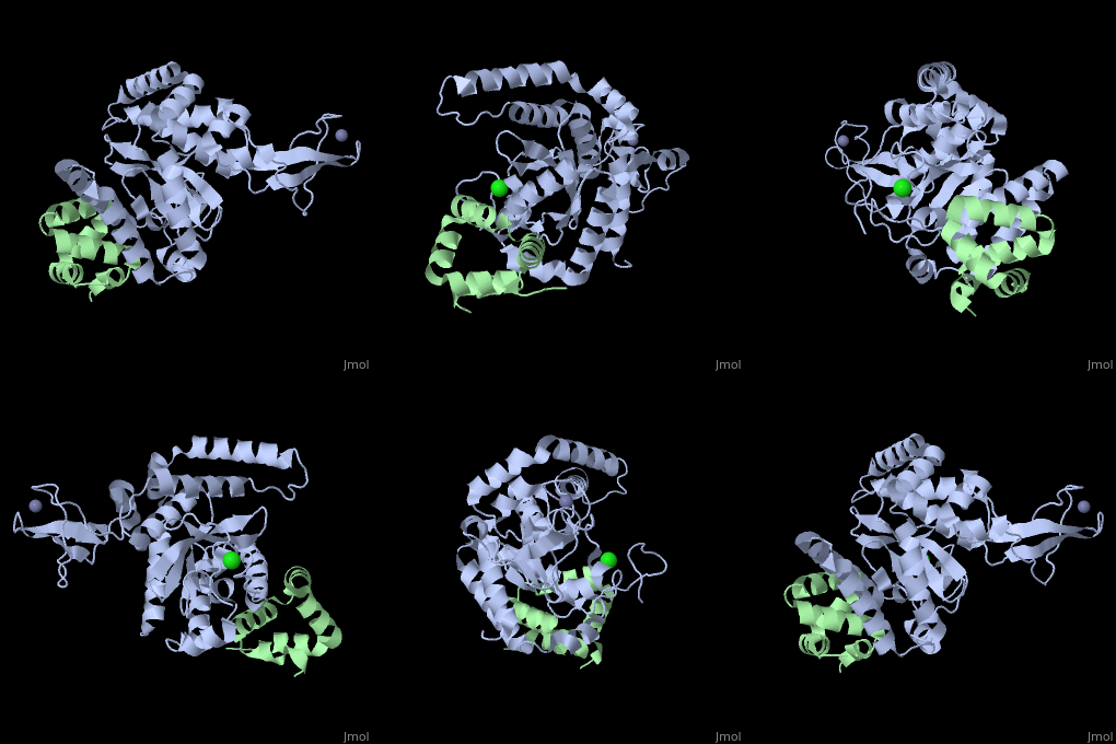
2f4m
From Proteopedia
| Line 4: | Line 4: | ||
|PDB= 2f4m |SIZE=350|CAPTION= <scene name='initialview01'>2f4m</scene>, resolution 1.85Å | |PDB= 2f4m |SIZE=350|CAPTION= <scene name='initialview01'>2f4m</scene>, resolution 1.85Å | ||
|SITE= | |SITE= | ||
| - | |LIGAND= <scene name='pdbligand= | + | |LIGAND= <scene name='pdbligand=CL:CHLORIDE+ION'>CL</scene>, <scene name='pdbligand=ZN:ZINC+ION'>ZN</scene> |
| - | |ACTIVITY= [http://en.wikipedia.org/wiki/Peptide-N(4)-(N-acetyl-beta-glucosaminyl)asparagine_amidase Peptide-N(4)-(N-acetyl-beta-glucosaminyl)asparagine amidase], with EC number [http://www.brenda-enzymes.info/php/result_flat.php4?ecno=3.5.1.52 3.5.1.52] | + | |ACTIVITY= <span class='plainlinks'>[http://en.wikipedia.org/wiki/Peptide-N(4)-(N-acetyl-beta-glucosaminyl)asparagine_amidase Peptide-N(4)-(N-acetyl-beta-glucosaminyl)asparagine amidase], with EC number [http://www.brenda-enzymes.info/php/result_flat.php4?ecno=3.5.1.52 3.5.1.52] </span> |
|GENE= Rad23b, Mhr23b ([http://www.ncbi.nlm.nih.gov/Taxonomy/Browser/wwwtax.cgi?mode=Info&srchmode=5&id=10090 Mus musculus]) | |GENE= Rad23b, Mhr23b ([http://www.ncbi.nlm.nih.gov/Taxonomy/Browser/wwwtax.cgi?mode=Info&srchmode=5&id=10090 Mus musculus]) | ||
| + | |DOMAIN= | ||
| + | |RELATEDENTRY=[[2f4o|2F4O]] | ||
| + | |RESOURCES=<span class='plainlinks'>[http://oca.weizmann.ac.il/oca-docs/fgij/fg.htm?mol=2f4m FirstGlance], [http://oca.weizmann.ac.il/oca-bin/ocaids?id=2f4m OCA], [http://www.ebi.ac.uk/pdbsum/2f4m PDBsum], [http://www.rcsb.org/pdb/explore.do?structureId=2f4m RCSB]</span> | ||
}} | }} | ||
| Line 29: | Line 32: | ||
[[Category: Zhao, G.]] | [[Category: Zhao, G.]] | ||
[[Category: Zhou, X.]] | [[Category: Zhou, X.]] | ||
| - | [[Category: CL]] | ||
| - | [[Category: ZN]] | ||
[[Category: glycoprotein]] | [[Category: glycoprotein]] | ||
[[Category: nucleotide excision repair]] | [[Category: nucleotide excision repair]] | ||
| Line 37: | Line 38: | ||
[[Category: ubiquitin-dependent protein degradation]] | [[Category: ubiquitin-dependent protein degradation]] | ||
| - | ''Page seeded by [http://oca.weizmann.ac.il/oca OCA ] on | + | ''Page seeded by [http://oca.weizmann.ac.il/oca OCA ] on Mon Mar 31 02:57:48 2008'' |
Revision as of 23:57, 30 March 2008
| |||||||
| , resolution 1.85Å | |||||||
|---|---|---|---|---|---|---|---|
| Ligands: | , | ||||||
| Gene: | Rad23b, Mhr23b (Mus musculus) | ||||||
| Activity: | Peptide-N(4)-(N-acetyl-beta-glucosaminyl)asparagine amidase, with EC number 3.5.1.52 | ||||||
| Related: | 2F4O
| ||||||
| Resources: | FirstGlance, OCA, PDBsum, RCSB | ||||||
| Coordinates: | save as pdb, mmCIF, xml | ||||||
The Mouse PNGase-HR23 Complex Reveals a Complete Remodulation of the Protein-Protein Interface Compared to its Yeast Orthologs
Overview
Peptide N-glycanase removes N-linked oligosaccharides from misfolded glycoproteins as part of the endoplasmic reticulum-associated degradation pathway. This process involves the formation of a tight complex of peptide N-glycanase with Rad23 in yeast and the orthologous HR23 proteins in mammals. In addition to its function in endoplasmic reticulum-associated degradation, HR23 is also involved in DNA repair, where it plays an important role in damage recognition in complex with the xeroderma pigmentosum group C protein. To characterize the dual role of HR23, we have determined the high resolution crystal structure of the mouse peptide N-glycanase catalytic core in complex with the xeroderma pigmentosum group C binding domain from HR23B. Peptide N-glycanase features a large cleft between its catalytic cysteine protease core and zinc binding domain. Opposite the zinc binding domain is the HR23B-interacting region, and surprisingly, the complex interface is fundamentally different from the orthologous yeast peptide N-glycanase-Rad23 complex. Different regions on both proteins are involved in complex formation, revealing an amazing degree of divergence in the interaction between two highly homologous proteins. Furthermore, the mouse peptide N-glycanase-HR23B complex mimics the interaction between xeroderma pigmentosum group C and HR23B, thereby providing a first structural model of how the two proteins interact within the nucleotide excision repair cascade in higher eukaryotes. The different interaction interfaces of the xeroderma pigmentosum group C binding domains in yeast and mammals suggest a co-evolution of the endoplasmic reticulum-associated degradation and DNA repair pathways.
About this Structure
2F4M is a Protein complex structure of sequences from Mus musculus. Full crystallographic information is available from OCA.
Reference
Structure of the mouse peptide N-glycanase-HR23 complex suggests co-evolution of the endoplasmic reticulum-associated degradation and DNA repair pathways., Zhao G, Zhou X, Wang L, Li G, Kisker C, Lennarz WJ, Schindelin H, J Biol Chem. 2006 May 12;281(19):13751-61. Epub 2006 Feb 24. PMID:16500903
Page seeded by OCA on Mon Mar 31 02:57:48 2008
Categories: Mus musculus | Peptide-N(4)-(N-acetyl-beta-glucosaminyl)asparagine amidase | Protein complex | Kisker, C. | Lennarz, W J. | Schindelin, H. | Wang, L. | Zhao, G. | Zhou, X. | Glycoprotein | Nucleotide excision repair | Peptide:n-glycanase | Transglutaminase | Ubiquitin-dependent protein degradation

