This old version of Proteopedia is provided for student assignments while the new version is undergoing repairs. Content and edits done in this old version of Proteopedia after March 1, 2026 will eventually be lost when it is retired in about June of 2026.
Apply for new accounts at the new Proteopedia. Your logins will work in both the old and new versions.
P53-DNA Recognition
From Proteopedia
| Line 75: | Line 75: | ||
A more general discussion of structural origins of binding specificity in protein-DNA recognition has been published along with a suggestion for a new '''classification of protein-DNA readout modes''' that goes beyond the historical description of direct and indirect readout<ref name="annualreview">Rohs R, Jin X, West SM, Joshi R, Honig B, Mann RS. Origins of specificity in protein-DNA recognition. Annu Rev Biochem. 2010;79:233-69. [http://www.ncbi.nlm.nih.gov/pubmed/20334529 PMID:20334529].</ref>.<br/> | A more general discussion of structural origins of binding specificity in protein-DNA recognition has been published along with a suggestion for a new '''classification of protein-DNA readout modes''' that goes beyond the historical description of direct and indirect readout<ref name="annualreview">Rohs R, Jin X, West SM, Joshi R, Honig B, Mann RS. Origins of specificity in protein-DNA recognition. Annu Rev Biochem. 2010;79:233-69. [http://www.ncbi.nlm.nih.gov/pubmed/20334529 PMID:20334529].</ref>.<br/> | ||
</StructureSection> | </StructureSection> | ||
| - | + | =3D structures of p53= | |
| + | [[P53]] | ||
=Acknowledgements= | =Acknowledgements= | ||
Revision as of 09:06, 31 December 2014
| |||||||||||
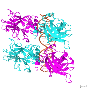


3D structures of p53
Acknowledgements
This Proteopedia page originates from the partnership of the Rohs Laboratory at the University of Southern California with La Cañada High School. This partnership was initiated by Remo Rohs and Patty Compeau in September 2011 as Bioinformatics Institute, which is part of the Institutes of the 21st Century. Advice and technical help by Proteopedia editors Eran Hodis, Eric Martz, Jaime Prilusky, and Joel Sussman is acknowledged.
References
- ↑ 1.0 1.1 1.2 1.3 Kitayner M, Rozenberg H, Rohs R, Suad O, Rabinovich D, Honig B, Shakked Z. Diversity in DNA recognition by p53 revealed by crystal structures with Hoogsteen base pairs. Nat Struct Mol Biol. 2010;17(4):423-9. PMID:20364130.
- ↑ Jeffrey PD, Gorina S, Pavletich NP. Crystal structure of the p53 tetramerization domain. Science 1995;267:1498-502. PMID:7878469.
- ↑ Kitayner M, Rozenberg H, Kessler N, Rabinovich D, Shaulov L, Haran TE, Shakked Z. Structural basis of DNA recognition by p53 tetramers. Mol Cell. 2006 Jun 23;22(6):741-53. PMID:16793544.
- ↑ Horvath MM, Wang X, Resnick MA, Bell DA. Divergent evolution of human p53 binding sites: cell cycle versus apoptosis. PLoS Genet. 2007 Jul;3(7):e127. PMID:17677004.
- ↑ Rohs R, West SM, Sosinsky A, Liu P, Mann RS, Honig B. The role of DNA shape in protein-DNA recognition. Nature. 2009;461(7268):1248-53. PMID:19865164.
- ↑ Aishima J, Gitti RK, Noah JE, Gan HH, Schlick T, Wolberger C. A Hoogsteen base pair embedded in undistorted B-DNA. Nucleic Acids Res. 2002;30(23):5244-52. PMID:12466549.
- ↑ Chen Y, Dey R, Chen L. Crystal structure of the p53 core domain bound to a full consensus site as a self-assembled tetramer. Structure. 2010;18(2):246-56. PMID:20159469.
- ↑ Nikolova EN, Kim E, Wise AA, O'Brien PJ, Andricioaei I, Al-Hashimi HM. Transient Hoogsteen base pairs in canonical duplex DNA. Nature. 2011;470(7335):498-502. PMID:21270796.
- ↑ Rohs R, Jin X, West SM, Joshi R, Honig B, Mann RS. Origins of specificity in protein-DNA recognition. Annu Rev Biochem. 2010;79:233-69. PMID:20334529.
Proteopedia Page Contributors and Editors (what is this?)
Remo Rohs, Eric Martz, Alexander Berchansky, Julia Tam, Sharon Kim, Bailey Holmes, Angel Herraez, Joseph M. Steinberger, Eran Hodis, Masha Karelina, Michal Harel, Ana Carolina Dantas Machado, Jaime Prilusky, Skyler Saleebyan, Joel L. Sussman, Keziah Kim
