We apologize for Proteopedia being slow to respond. For the past two years, a new implementation of Proteopedia has been being built. Soon, it will replace this 18-year old system. All existing content will be moved to the new system at a date that will be announced here.
Sandbox Reserved 970
From Proteopedia
(Difference between revisions)
| Line 3: | Line 3: | ||
== Introduction == | == Introduction == | ||
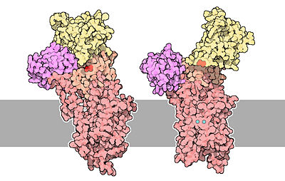
| - | P-type ATPase pump | + | All living organisms depend on P-type ATPase to pump cation across the membrane. P type ATPase are distinct from other ATPases beecause there is an autophosphorylation step in the catalytic cycle. They play a fundamental role in organisms metabolism and physiology. Ca2+ ATPase is one type of P-type ATPase which transport calcium ions across the membrane against a concentration gradient. These pumps clear cytoplasm of the second messenger, the calcium. It's very important to keep a low concentration of calcium in the cell for a good cell signaling. The hydrolysis of ATP is necessary for the pump's functioning. For each ATP hydrolysed, it transfers two calcium ions through the membrane, and two or three hydrogen ions in the opposite direction. |
| - | + | ||
| - | {{clear}} | ||
== Structural Highlights == | == Structural Highlights == | ||
| Line 17: | Line 16: | ||
| - | == | + | == Mechanism of action == |
The architecture of calcium ATPase (determined by X-Ray crystallography) allow to understand mechanisms by which the energy of ATP is coupled to the calcium transport across a membrane. | The architecture of calcium ATPase (determined by X-Ray crystallography) allow to understand mechanisms by which the energy of ATP is coupled to the calcium transport across a membrane. | ||
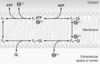
| - | The first step of the calcium pump catalytic cycle is the cooperative binding of <scene name='60/604489/Calcium_molecules/1'>two calcium ions</scene> in the calcium binding cavity. Then, ATP binds to the ATP binding site (nucleotide binding domain) and transfers its γ-phosphate to the <scene name='60/604489/Asp_351/1'>aspartic acide 351</scene> (phosphorylation domain). That creates a acid-stable aspartyl phosphate intermediate. The phosphorylation of Asp351 allows a large conformational changes in cytoplasmic domains: the nucleotide binding domain and the phosphorylation domain are brought into close proximity. This rearrangement causes a 90° rotation of the actuator domain, which leads to a rearrangement of the transmembrane helices. This rearrangement alters the affinity of the protein for the calcium and disrupts the calcium binding cavity. Calcium is released in the lumen of the endoplasmic reticulum/Golgi Apparatus or outside the cell. After releasing calcium, two or three protons bind to the transport sites (charges compensation) and the aspartyl phosphate is hydrolyzed to complete the cycle. <ref>Marisa Brini , Ernesto Carafoli, 2009 - ''Calcium pumps in health and disease'' - Physiological Reviews</ref> | + | The first step of the calcium pump catalytic cycle is the cooperative binding of <scene name='60/604489/Calcium_molecules/1'>two calcium ions</scene> in the calcium binding cavity. Then, ATP binds to the ATP binding site (nucleotide binding domain) and transfers its γ-phosphate to the <scene name='60/604489/Asp_351/1'>aspartic acide 351</scene> (phosphorylation domain). That creates a acid-stable aspartyl phosphate intermediate. The phosphorylation of Asp351 allows a large conformational changes in cytoplasmic domains: the nucleotide binding domain and the phosphorylation domain are brought into close proximity. This rearrangement causes a 90° rotation of the actuator domain, which leads to a rearrangement of the transmembrane helices. This rearrangement alters the affinity of the protein for the calcium and disrupts the calcium binding cavity. Calcium is released in the lumen of the endoplasmic reticulum/Golgi Apparatus or outside the cell. After releasing calcium, two or three protons bind to the transport sites (charges compensation) and the aspartyl phosphate is hydrolyzed to complete the cycle. <ref name = "four">Marisa Brini , Ernesto Carafoli, 2009 - ''Calcium pumps in health and disease'' - Physiological Reviews</ref> |
[[Image:51-TheCalciumPumps-calcium-pumps.jpg|400px|center|]] | [[Image:51-TheCalciumPumps-calcium-pumps.jpg|400px|center|]] | ||
| Line 28: | Line 27: | ||
| - | To sum up, calcium pumps have two conformations, E1 and E2. These two conformations are characterized by different specificity for ion binding. When the pump is in the E1 state, it has high calcium affinity and interacts with calcium at one side of the membrane. In the E2 state, the enzyme has a lower calcium affinity and that leads to the release of the ion at the opposite side. E1 has the calcium binding site oriented toward the cytoplasm. E2 has the calcium binding site oriented toward the lumen of the endoplasmic reticulum or toward the extracellular background<ref name="third">Thomas D.Pollard and William C. Earnshaw, - ''Membrane, structure and function'' - Cell Biology (second edition), p.133-136</ref>. The phosphorylated intermediate, E1 can phosphorylate ADP, whereas E2 can only react with water<ref>David H.MacLennan, William J.Rice and N. Michael Green, 1997 - ''The Mechanism of Ca2+ Transport by Sarco(Endo)plasmic Reticulum Ca2+-ATPases'' - The Journal of Biological Chemistry, p.272, 28815-28818, http://www.jbc.org/content/272/46/28815.full.html</ref>. | + | To sum up, calcium pumps have two conformations, E1 and E2. These two conformations are characterized by different specificity for ion binding. When the pump is in the E1 state, it has high calcium affinity and interacts with calcium at one side of the membrane. In the E2 state, the enzyme has a lower calcium affinity and that leads to the release of the ion at the opposite side. E1 has the calcium binding site oriented toward the cytoplasm. E2 has the calcium binding site oriented toward the lumen of the endoplasmic reticulum or toward the extracellular background<ref name="third">Thomas D.Pollard and William C. Earnshaw, - ''Membrane, structure and function'' - Cell Biology (second edition), p.133-136</ref>. The phosphorylated intermediate, E1 can phosphorylate ADP, whereas E2 can only react with water<ref>David H.MacLennan, William J.Rice and N. Michael Green, 1997 - ''The Mechanism of Ca2+ Transport by Sarco(Endo)plasmic Reticulum Ca2+-ATPases'' - The Journal of Biological Chemistry, p.272, 28815-28818, http://www.jbc.org/content/272/46/28815.full.html</ref>. In the absence of calcium, calcium pumps are mainly in E1 state. |
| - | [[Image:Etat.jpg|center|]] | + | [[Image:Etat.jpg|center|]]<ref name = "four">Marisa Brini , Ernesto Carafoli, 2009 - ''Calcium pumps in health and disease'' - Physiological Reviews</ref> |
| - | + | ||
| Line 41: | Line 39: | ||
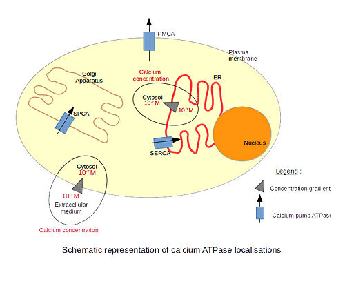
There are 3 types of calcium ATPase depending on their localization. The '''SERCA''' (Sarco/Endoplasmic Reticulum Calcium ATPase) is localized in the endoplasmic reticulum membranes, including the nuclear envelope. Only the structure of the SERCA has been resolved by x-ray crystallography. The '''PMCA''' (Plasma Membrane Calcium ATPase) is localized in plasma membranes. The '''SPCA''' (Secretory Pathway Calcium ATPase) is localized in the Golgi apparatus membranes. The particularity of this pump is that it is also able to transport Mn2+ ions. | There are 3 types of calcium ATPase depending on their localization. The '''SERCA''' (Sarco/Endoplasmic Reticulum Calcium ATPase) is localized in the endoplasmic reticulum membranes, including the nuclear envelope. Only the structure of the SERCA has been resolved by x-ray crystallography. The '''PMCA''' (Plasma Membrane Calcium ATPase) is localized in plasma membranes. The '''SPCA''' (Secretory Pathway Calcium ATPase) is localized in the Golgi apparatus membranes. The particularity of this pump is that it is also able to transport Mn2+ ions. | ||
| - | [[Image: | + | [[Image:ATPases.jpg|500px|center|]] |
Calcium is a very important molecule for cell signalling. Eucaryotic cells need to maintain a very low calcium concentration in their cytosol. The extracellular calcium concentration is much higher. That difference of concentrations across the membrane creates a gradient of concentration and allows the signalling to be very effective. Indeed, even a very small influx of calcium significantly increases the concentration of calcium inside the cell. Therefore, calcium pumps are very important to maintain the calcium concentration gradient and to remove calcium from the cell after signalling. | Calcium is a very important molecule for cell signalling. Eucaryotic cells need to maintain a very low calcium concentration in their cytosol. The extracellular calcium concentration is much higher. That difference of concentrations across the membrane creates a gradient of concentration and allows the signalling to be very effective. Indeed, even a very small influx of calcium significantly increases the concentration of calcium inside the cell. Therefore, calcium pumps are very important to maintain the calcium concentration gradient and to remove calcium from the cell after signalling. | ||
Calcium is involved in many physiological processes such as programmed cell death, fertilization, gene transcription, secretion (including neurotransmitter secretion) etc. For example, the SERCA pump is mainly found in skeletal muscle cells and cardiac cells. It is involved in the relaxation of skeletal muscle cells. Those cells contain a special endoplasmic reticulum called the sarcoplasmic reticulum which is the place of calcium storage. After contraction, calcium ions are transported from the cytoplasm into the sarcoplasmic reticulum through the SERCA pump. This causes the relaxation of the muscle cells because the cytosolic concentration of calcium decreases. The SERCA pump works together with the PMCA pump to export calcium ions from the cytosol and to set the resting level of the cytosolic calcium concentration<ref name="first">Benjamin Lewin, 2007 - Cells - Jones & Bartlett Learning</ref>. | Calcium is involved in many physiological processes such as programmed cell death, fertilization, gene transcription, secretion (including neurotransmitter secretion) etc. For example, the SERCA pump is mainly found in skeletal muscle cells and cardiac cells. It is involved in the relaxation of skeletal muscle cells. Those cells contain a special endoplasmic reticulum called the sarcoplasmic reticulum which is the place of calcium storage. After contraction, calcium ions are transported from the cytoplasm into the sarcoplasmic reticulum through the SERCA pump. This causes the relaxation of the muscle cells because the cytosolic concentration of calcium decreases. The SERCA pump works together with the PMCA pump to export calcium ions from the cytosol and to set the resting level of the cytosolic calcium concentration<ref name="first">Benjamin Lewin, 2007 - Cells - Jones & Bartlett Learning</ref>. | ||
| - | + | ||
== Regulations == | == Regulations == | ||
| - | There are different kind of calcium ATPase regulations. For example, the phospholamban (PLN or PLB) | + | There are different kind of calcium ATPase regulations. For example, the phospholamban (PLN or PLB) and the sarcolipin are membrane proteins that regulate the calcium pump in cardiac muscle and skeletal muscle cells. These two proteins are close homologous and play the same role. The phospholamban is a phosphoprotein that can be phosphorylated at three distinct sites by various protein kinases (PKA, PKC, CamK...). The phosphorylation state is mediated through beta-adrenergic stimulation. In unphosphorylate state, the phospholamban inhibits the activity of calcium pump in cardiac and skeletal muscle cells by decreasing the apparent affinity of the ATPase for calcium. The phosphorylation of the protein results in the dissociation of the protein from the ATPase. The phosphoprotein binds just downstream of the asp351 residue. |
Most of the activation mechanisms implicate the C-terminal region of the pump containing the high affinity calmodulin binding domain, which is involved in the autoinhibition of the pump<ref>Marianela G.Dalghi, Marisa M.Fernández, Mariela Ferreira-Gomes, Irene C.Mangialavori, Emilio L.Malchiodi, Emanuel E.Strehler and Juan Pablo F.C.Rossi, 2013 - ''Plasma Membrane Calcium ATPase Activity Is Regulated by Actin Oligomers through Direct Interaction'' - The Journal of Biological Chemistry, p.288, 23380-23393, http://www.jbc.org/content/288/32/23380.full.</ref>. | Most of the activation mechanisms implicate the C-terminal region of the pump containing the high affinity calmodulin binding domain, which is involved in the autoinhibition of the pump<ref>Marianela G.Dalghi, Marisa M.Fernández, Mariela Ferreira-Gomes, Irene C.Mangialavori, Emilio L.Malchiodi, Emanuel E.Strehler and Juan Pablo F.C.Rossi, 2013 - ''Plasma Membrane Calcium ATPase Activity Is Regulated by Actin Oligomers through Direct Interaction'' - The Journal of Biological Chemistry, p.288, 23380-23393, http://www.jbc.org/content/288/32/23380.full.</ref>. | ||
Revision as of 19:54, 7 January 2015
| |||||||||||
References
- ↑ 1.0 1.1 Benjamin Lewin, 2007 - Cells - Jones & Bartlett Learning
- ↑ 2.0 2.1 Marisa Brini , Ernesto Carafoli, 2009 - Calcium pumps in health and disease - Physiological Reviews
- ↑ David Goodsell, 2004 - Calcium pump molecul of the month - PDB, doi: 10.2210/rcsb_pdb/mom_2004_3
- ↑ Thomas D.Pollard and William C. Earnshaw, - Membrane, structure and function - Cell Biology (second edition), p.133-136
- ↑ David H.MacLennan, William J.Rice and N. Michael Green, 1997 - The Mechanism of Ca2+ Transport by Sarco(Endo)plasmic Reticulum Ca2+-ATPases - The Journal of Biological Chemistry, p.272, 28815-28818, http://www.jbc.org/content/272/46/28815.full.html
- ↑ Marianela G.Dalghi, Marisa M.Fernández, Mariela Ferreira-Gomes, Irene C.Mangialavori, Emilio L.Malchiodi, Emanuel E.Strehler and Juan Pablo F.C.Rossi, 2013 - Plasma Membrane Calcium ATPase Activity Is Regulated by Actin Oligomers through Direct Interaction - The Journal of Biological Chemistry, p.288, 23380-23393, http://www.jbc.org/content/288/32/23380.full.
- ↑ Marisa Brini and Ernesto Carafoli, 2010 - The plasma membrane Ca2+ ATPase and the Plasma Membrane Sodium Calcium Exchanger Cooperate in the Regulation of Cell Calcium - Cold Spring Harbor Perspectives in Biology, http://cshperspectives.cshlp.org/content/3/2/a004168.full


