We apologize for Proteopedia being slow to respond. For the past two years, a new implementation of Proteopedia has been being built. Soon, it will replace this 18-year old system. All existing content will be moved to the new system at a date that will be announced here.
Z-DNA
From Proteopedia
(Difference between revisions)
| Line 2: | Line 2: | ||
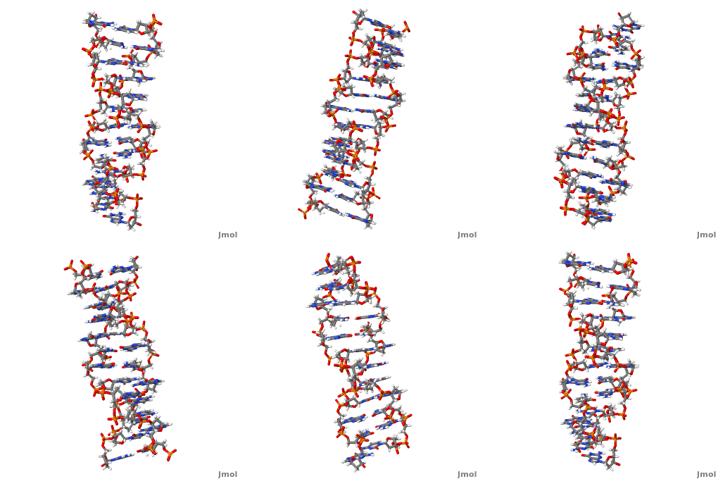
'''Z-DNA''' <scene name='Z-DNA/Z-dna_new/1'>(default scene)</scene> is a form of DNA that has a different structure from the more common <scene name='Sandbox_Z-DNA/Bdna/3'>B-DNA</scene> form.It is a left-handed double helix wherein the sugar-phosphate backbone has a zigzag pattern due to the alternate stacking of bases in [http://proteopedia.org/wiki/index.php/Syn_and_anti_nucleosides anti-conformation and syn conformation]. In Z-DNA only a minor groove is present and the major groove is absent. The residues that allow sequence-specific recognition of Z-DNA are present on the convex outer surface.<ref name = 'Rich'> PMID:12838348</ref> This DNA form is thought to play a role in the regulation of gene expression, DNA processing events and/or genetic instability.<ref name = 'Wang'>PMID:17485386</ref> | '''Z-DNA''' <scene name='Z-DNA/Z-dna_new/1'>(default scene)</scene> is a form of DNA that has a different structure from the more common <scene name='Sandbox_Z-DNA/Bdna/3'>B-DNA</scene> form.It is a left-handed double helix wherein the sugar-phosphate backbone has a zigzag pattern due to the alternate stacking of bases in [http://proteopedia.org/wiki/index.php/Syn_and_anti_nucleosides anti-conformation and syn conformation]. In Z-DNA only a minor groove is present and the major groove is absent. The residues that allow sequence-specific recognition of Z-DNA are present on the convex outer surface.<ref name = 'Rich'> PMID:12838348</ref> This DNA form is thought to play a role in the regulation of gene expression, DNA processing events and/or genetic instability.<ref name = 'Wang'>PMID:17485386</ref> | ||
| + | |||
| + | See also [[Z-DNA model tour]]. | ||
== Structure == | == Structure == | ||
| - | Z-DNA (<scene name='Sandbox_Z-DNA/B-z/7'>B-Z DNA junction</scene>, PDB entry [[2acj]]) can form '' | + | Z-DNA (<scene name='Sandbox_Z-DNA/B-z/7'>B-Z DNA junction</scene>, PDB entry [[2acj]]) can form ''in vitro'' from B-DNA by raising negative super helical stress or under low salt conditions when deoxycytosine is 5-methylated. The formation of Z-DNA ''in vivo'' is an energy requiring process. It forms behind a RNA polymerase moving through a DNA double helix during transcription and is subsequently stabilized due to the generation of negative supercoils. Z-DNA is the first single crystal X-ray structure of a DNA fragment. It was crystallized as a self complementary DNA hexamer d(CG)<sub>3</sub> by Andrew Wang, Alexander Rich and their co-workers at MIT in 1979. <ref name = 'Rich'>PMID:12838348</ref><ref name ='Wang'>PMID:17485386</ref> |
Whenever B-DNA transforms into Z-DNA two <scene name='Sandbox_Z-DNA/B-zjunction/7'>B-Z junctions</scene> form. The crystal structure of these junctions revealed<scene name='Sandbox_Z-DNA/Extruded/12'> two extruded bases</scene>, <scene name='Z-DNA/Extruded/2'>adenine</scene> and <scene name='Z-DNA/Extruded/3'>thymine</scene> at the junction. A crucial finding from this structure is that a right handed DNA can transform to a left handed DNA or vice versa by the disruption and extrusion of a base pair. It has also been suggested that the extruded base pairs at B-Z DNA junction may be sites for DNA modification.<ref>PMID:16237447</ref> | Whenever B-DNA transforms into Z-DNA two <scene name='Sandbox_Z-DNA/B-zjunction/7'>B-Z junctions</scene> form. The crystal structure of these junctions revealed<scene name='Sandbox_Z-DNA/Extruded/12'> two extruded bases</scene>, <scene name='Z-DNA/Extruded/2'>adenine</scene> and <scene name='Z-DNA/Extruded/3'>thymine</scene> at the junction. A crucial finding from this structure is that a right handed DNA can transform to a left handed DNA or vice versa by the disruption and extrusion of a base pair. It has also been suggested that the extruded base pairs at B-Z DNA junction may be sites for DNA modification.<ref>PMID:16237447</ref> | ||
Revision as of 21:13, 11 January 2017
| |||||||||||
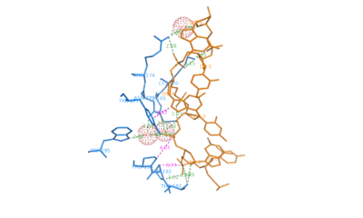
Movie Depicting ADAR1 binding to Z-DNA
Comparison of the three helices and helical parameters of DNA
Sources[7]
|
|
|
| Parameter | A-DNA | B-DNA | Z-DNA |
|---|---|---|---|
| Helix sense | right-handed | right-handed | left-handed |
| Residues per turn | 11 | 10.5 | 12 |
| Axial rise [Å] | 2.55 | 3.4 | 3.7 |
| Helix pitch(°) | 28 | 34 | 45 |
| Base pair tilt(°) | 20 | −6 | 7 |
| Rotation per residue (°) | 33 | 36 | -30 |
| Diameter of helix [Å] | 23 | 20 | 18 |
| Glycosidic bond configuration dA,dT,dC dG | anti anti | anti anti | anti syn |
| Sugar pucker dA,dT,dC dG | C3'-endo C3'-endo | C2'-endo C2'-endo | C2'-endo C3'-endo |
| Intrastrand phosphate-phosphate distance [Å] dA,dT,dC dG | 5.9 5.9 | 7.0 7.0 | 7.0 5.9 |
| Sources:[8][9][10] | |||
3D structures of Z-DNA
Updated on 11-January-2017
1woe, 3qba – ZDNA hexamer CGCGCG
1qbj – ZDNA hexamer CGCGCG + hADAR1 Zα domain - human
2heo - ZDNA hexamer CGCGCG + Z-DNA-binding protein Zα domain – mouse
2eyi - ZDNA hexamer CGCGCG + Z-DNA-binding protein Zβ domain
1j75 - ZDNA hexamer CGCGCG + DLM-1 Zα domain
Additional Resources
For additional information, see: Nucleic Acids
References
- ↑ 1.0 1.1 1.2 Rich A, Zhang S. Timeline: Z-DNA: the long road to biological function. Nat Rev Genet. 2003 Jul;4(7):566-72. PMID:12838348 doi:10.1038/nrg1115
- ↑ 2.0 2.1 2.2 2.3 2.4 2.5 Wang G, Vasquez KM. Z-DNA, an active element in the genome. Front Biosci. 2007 May 1;12:4424-38. PMID:17485386
- ↑ Ha SC, Lowenhaupt K, Rich A, Kim YG, Kim KK. Crystal structure of a junction between B-DNA and Z-DNA reveals two extruded bases. Nature. 2005 Oct 20;437(7062):1183-6. PMID:16237447 doi:10.1038/nature04088
- ↑ Schwartz T, Rould MA, Lowenhaupt K, Herbert A, Rich A. Crystal structure of the Zalpha domain of the human editing enzyme ADAR1 bound to left-handed Z-DNA. Science. 1999 Jun 11;284(5421):1841-5. PMID:10364558
- ↑ Kim YG, Lowenhaupt K, Oh DB, Kim KK, Rich A. Evidence that vaccinia virulence factor E3L binds to Z-DNA in vivo: Implications for development of a therapy for poxvirus infection. Proc Natl Acad Sci U S A. 2004 Feb 10;101(6):1514-8. Epub 2004 Feb 2. PMID:14757814 doi:10.1073/pnas.0308260100
- ↑ Drozdzal P, Gilski M, Kierzek R, Lomozik L, Jaskolski M. High-resolution crystal structure of Z-DNA in complex with Cr cations. J Biol Inorg Chem. 2015 Feb 17. PMID:25687556 doi:http://dx.doi.org/10.1007/s00775-015-1247-5
- ↑ http://203.129.231.23/indira/nacc/
- ↑ Rich A, Nordheim A, Wang AH. The chemistry and biology of left-handed Z-DNA. Annu Rev Biochem. 1984;53:791-846. PMID:6383204 doi:http://dx.doi.org/10.1146/annurev.bi.53.070184.004043
- ↑ Wang AH, Quigley GJ, Kolpak FJ, Crawford JL, van Boom JH, van der Marel G, Rich A. Molecular structure of a left-handed double helical DNA fragment at atomic resolution. Nature. 1979 Dec 13;282(5740):680-6. PMID:514347
- ↑ Sinden, Richard R (1994-01-15). DNA structure and function (1st ed.). Academic Press. pp. 398. ISBN 0-12-645750-6.
Proteopedia Page Contributors and Editors (what is this?)
Adithya Sagar, Michal Harel, Joel L. Sussman, Alexander Berchansky, David Canner, Donald Voet, Jaime Prilusky, Karl Oberholser, Eran Hodis


