We apologize for Proteopedia being slow to respond. For the past two years, a new implementation of Proteopedia has been being built. Soon, it will replace this 18-year old system. All existing content will be moved to the new system at a date that will be announced here.
Sandbox Reserved 1075
From Proteopedia
(Difference between revisions)
| Line 2: | Line 2: | ||
<StructureSection load='4W4I' size='340' side='right' caption='Caption for this structure' scene=''> | <StructureSection load='4W4I' size='340' side='right' caption='Caption for this structure' scene=''> | ||
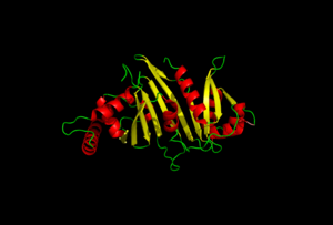
| - | EspG secretion is key in understanding the virulence of ''mycobacterium tuberculosis''. | + | EspG secretion is key in understanding the virulence of ''mycobacterium tuberculosis''. The specificity of EspG binding affinity to its specific PE-PPE ligand have many contributing factors. The four different EspG proteins found in ''mycobacterium tuberculosis'' have different characteristics that influence binding, where EspG5 binds to the most PE-PPE proteins. |
You may include any references to papers as in: the use of JSmol in Proteopedia <ref>DOI 10.1002/ijch.201300024</ref> or to the article describing Jmol <ref>PMID:21638687</ref> to the rescue. | You may include any references to papers as in: the use of JSmol in Proteopedia <ref>DOI 10.1002/ijch.201300024</ref> or to the article describing Jmol <ref>PMID:21638687</ref> to the rescue. | ||
| Line 12: | Line 12: | ||
=== Excretion === | === Excretion === | ||
| + | EspG PE-PPE excretion is done through the ESX secretion pathway. | ||
| + | |||
=== Binding === | === Binding === | ||
| + | Factors that influence binding of EspG and PE-PPE: | ||
| + | electrostatics | ||
| + | shape | ||
| + | ligand random coil | ||
| + | key catalytic residues | ||
| + | |||
=== Pocket Residues === | === Pocket Residues === | ||
| + | Jon | ||
== Structural highlights == | == Structural highlights == | ||
| + | |||
| + | |||
== Relevance == | == Relevance == | ||
| - | <scene name='69/697501/Helix_highlited/1'>Helix Highlited</scene> | ||
| - | This is a sample scene created with SAT to <scene name="/12/3456/Sample/1">color</scene> by Group, and another to make <scene name="/12/3456/Sample/2">a transparent representation</scene> of the protein. You can make your own scenes on SAT starting from scratch or loading and editing one of these sample scenes. | ||
| Line 35: | Line 44: | ||
| + | |||
| + | <scene name='69/697501/Helix_highlited/1'>Helix Highlited</scene> | ||
| + | |||
| + | This is a sample scene created with SAT to <scene name="/12/3456/Sample/1">color</scene> by Group, and another to make <scene name="/12/3456/Sample/2">a transparent representation</scene> of the protein. You can make your own scenes on SAT starting from scratch or loading and editing one of these sample scenes. | ||
== References == | == References == | ||
<references/> | <references/> | ||
Revision as of 12:46, 24 March 2015
Binding Specificity of EspG Proteins to PE-PPE Proteins in mycobacterium tuberculosis
| |||||||||||
This is a sample scene created with SAT to by Group, and another to make of the protein. You can make your own scenes on SAT starting from scratch or loading and editing one of these sample scenes.
References
- ↑ Hanson, R. M., Prilusky, J., Renjian, Z., Nakane, T. and Sussman, J. L. (2013), JSmol and the Next-Generation Web-Based Representation of 3D Molecular Structure as Applied to Proteopedia. Isr. J. Chem., 53:207-216. doi:http://dx.doi.org/10.1002/ijch.201300024
- ↑ Herraez A. Biomolecules in the computer: Jmol to the rescue. Biochem Mol Biol Educ. 2006 Jul;34(4):255-61. doi: 10.1002/bmb.2006.494034042644. PMID:21638687 doi:10.1002/bmb.2006.494034042644
- ↑ Ekiert DC, Cox JS. Structure of a PE-PPE-EspG complex from Mycobacterium tuberculosis reveals molecular specificity of ESX protein secretion. Proc Natl Acad Sci U S A. 2014 Oct 14;111(41):14758-63. doi:, 10.1073/pnas.1409345111. Epub 2014 Oct 1. PMID:25275011 doi:http://dx.doi.org/10.1073/pnas.1409345111

