We apologize for Proteopedia being slow to respond. For the past two years, a new implementation of Proteopedia has been being built. Soon, it will replace this 18-year old system. All existing content will be moved to the new system at a date that will be announced here.
Sandbox Reserved 1058
From Proteopedia
(Difference between revisions)
| Line 7: | Line 7: | ||
Isocitrate lyase plays a key role in survival of ''M. tuberculosis'' by sustaining intracellular infections in inflammatory respiratory macrophages. Used in the citric acid cycle, isocitrate lyase is the first enzyme catalyzing the carbon conserving glyoxylate pathway. This glyoxylate pathway has not been observed in mammals and thus presents a unique drug target to solely attack TB infections. | Isocitrate lyase plays a key role in survival of ''M. tuberculosis'' by sustaining intracellular infections in inflammatory respiratory macrophages. Used in the citric acid cycle, isocitrate lyase is the first enzyme catalyzing the carbon conserving glyoxylate pathway. This glyoxylate pathway has not been observed in mammals and thus presents a unique drug target to solely attack TB infections. | ||
===Mechanism of Action=== | ===Mechanism of Action=== | ||
| - | + | [[Image:TCA_Cycle.png|250 px|center|thumb|'''Figure 1. Citric Acid Cycle with Glyoxylate Shunt Pathway.''' In several bacterial species, there is a carbon conserving gloxylate shunt pathway that converts isocitrate to malate in two steps instead of the usual five steps.]] | |
==Protein Structure== | ==Protein Structure== | ||
===Crystal Structure=== | ===Crystal Structure=== | ||
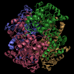
| - | [[Image:Normal_Crystal_Structure.png|250 px|center|thumb|'''Figure | + | [[Image:Normal_Crystal_Structure.png|250 px|center|thumb|'''Figure 2. Structure of Isocitrate Lyase.''' Quaternary structure is comprised of four subunits forming an alpha/beta barrel.]] |
Isocitrate lyase (PDB Code 1F8I) is a tetramer with 222 symmetry. Each subunit is composed of 14 alpha helices and 14 beta sheets. A unique structural feature of this enzyme is a phenomenon called "<scene name='69/694225/Helix_swapping/1'>helix swapping</scene>". | Isocitrate lyase (PDB Code 1F8I) is a tetramer with 222 symmetry. Each subunit is composed of 14 alpha helices and 14 beta sheets. A unique structural feature of this enzyme is a phenomenon called "<scene name='69/694225/Helix_swapping/1'>helix swapping</scene>". | ||
Helix swapping is observed between two monomers to form stable dimers. The 11th and 12th helices of each monomer exchange three dimensional placement with the respective helices of the opposite monomer. Due to the 222 symmetry observed, only two dimers form than combine to form the observed tetramer. As a result of this structure, 18% of the surface of each monomer is buried within the protein. | Helix swapping is observed between two monomers to form stable dimers. The 11th and 12th helices of each monomer exchange three dimensional placement with the respective helices of the opposite monomer. Due to the 222 symmetry observed, only two dimers form than combine to form the observed tetramer. As a result of this structure, 18% of the surface of each monomer is buried within the protein. | ||
===Ligand Bound=== | ===Ligand Bound=== | ||
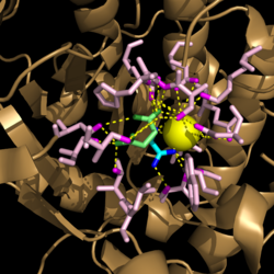
| - | [[Image:Active_Site_Hydrogen_Bonding.png|250 px|center|thumb|'''Figure | + | [[Image:Active_Site_Hydrogen_Bonding.png|250 px|center|thumb|'''Figure 3. Active site residues hydrogen bound to a cofactor and the products of the catalyzed isocitrate reaction.''' Glyoxylate is shown in blue, succinate is shown in green, and the Mg<sup>2+</sup> cofactor is shown in yellow.]] |
== Function == | == Function == | ||
Revision as of 17:35, 7 April 2015
Isocitrate Lyase from Mycobacterium tuberculosis
| |||||||||||


