Sandbox Reserved 1075
From Proteopedia
(Difference between revisions)
| Line 12: | Line 12: | ||
Through specific binding factors, an EspG binds to its PE-PPE ligand to be secreted through the ESAT pathway. Though the ESAT-6 secretion system is poorly understood, it is known that PE-PPE proteins and EspG proteins influence virulence and pathogenicity of the infection. | Through specific binding factors, an EspG binds to its PE-PPE ligand to be secreted through the ESAT pathway. Though the ESAT-6 secretion system is poorly understood, it is known that PE-PPE proteins and EspG proteins influence virulence and pathogenicity of the infection. | ||
| - | + | ||
| + | == Structural Differences == | ||
| + | |||
| + | [[Image:EspG5_and_EspG3_overlap_W.png|250 px|left|thumb|EspG5 and EspG3]] | ||
| + | |||
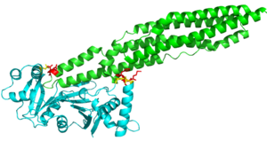
| + | Here are the key differences found on the tertiary structure of EspG3 (yellow) with EspG5(blue). The highlighted alpha helix shows a difference in length between EspG3 and EspG5. This difference in length contributes to the steric hinderance when binding to a PE-PPE ligand. The random loop highlighted toward the bottom of this protein varies in length between EspG proteins, this influences binding. The small random turn highlighted shows inconceivable difference to the EspG5 protein. The β-2, β-3 sequence similarity between EspG5 and EspG3 is very low. | ||
| + | |||
== Excretion == | == Excretion == | ||
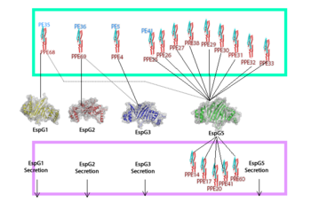
EspG PE-PPE excretion is done through the ESX secretion pathway. Mycobacterium tuberculosis uses this ESX-1 secretion system to deliver virulence factors into host macrophage and monocyte white blood cells during the 'Mycobacterium tuberculosis' infection. Though the exact excreted protein is currently unknown, several different PE-PPE complexes and EspG complexes have been identified. In the image "Specificity of EspG Binding", it can be noted how the specificity of specific PE-PPE complexes only bind to certain EspG proteins. To exemplify this, notice the factors that influence binding in EspG5 with PE25-PPE41 and see the contrast when EspG3 attempts PE25-PPE41. <ref>PMID:15973432</ref> | EspG PE-PPE excretion is done through the ESX secretion pathway. Mycobacterium tuberculosis uses this ESX-1 secretion system to deliver virulence factors into host macrophage and monocyte white blood cells during the 'Mycobacterium tuberculosis' infection. Though the exact excreted protein is currently unknown, several different PE-PPE complexes and EspG complexes have been identified. In the image "Specificity of EspG Binding", it can be noted how the specificity of specific PE-PPE complexes only bind to certain EspG proteins. To exemplify this, notice the factors that influence binding in EspG5 with PE25-PPE41 and see the contrast when EspG3 attempts PE25-PPE41. <ref>PMID:15973432</ref> | ||
| Line 46: | Line 52: | ||
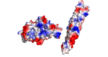
'''Electrostatics:''' | '''Electrostatics:''' | ||
There is an overall negative charge on the PE-PPE complex, and the binding tip is only partially negative. On the EspG5 protein, the binding pocket is partially positive, which aids coupling of the EspG & PE-PPE complex. The other EspG proteins found in 'Mycobacterium tuberculosis' have different electrostatic pocket charges which prevent binding of the PE25-PPE41 ligand. | There is an overall negative charge on the PE-PPE complex, and the binding tip is only partially negative. On the EspG5 protein, the binding pocket is partially positive, which aids coupling of the EspG & PE-PPE complex. The other EspG proteins found in 'Mycobacterium tuberculosis' have different electrostatic pocket charges which prevent binding of the PE25-PPE41 ligand. | ||
| - | |||
[[Image:EspG5_Electrostatics_W.png|350 px|left|thumb|Electrostatics]] | [[Image:EspG5_Electrostatics_W.png|350 px|left|thumb|Electrostatics]] | ||
| - | |||
| - | == Structural highlights == | ||
| - | |||
| - | [[Image:EspG5_and_EspG3_overlap_W.png|250 px|left|thumb|EspG5 and EspG3]] | ||
| - | |||
| - | Here are the key differences found on the tertiary structure of EspG3 (yellow) with EspG5(blue). The highlighted alpha helix shows a difference in length between EspG3 and EspG5. This difference in length contributes to the steric hinderance when binding to a PE-PPE ligand. The random loop highlighted toward the bottom of this protein varies in length between EspG proteins, this influences binding. The small random turn highlighted shows inconceivable difference to the EspG5 protein. The β-2, β-3 sequence similarity between EspG5 and EspG3 is very low. | ||
| - | |||
Revision as of 01:03, 16 April 2015
| |||||||||||
References
- ↑ Ekiert DC, Cox JS. Structure of a PE-PPE-EspG complex from Mycobacterium tuberculosis reveals molecular specificity of ESX protein secretion. Proc Natl Acad Sci U S A. 2014 Oct 14;111(41):14758-63. doi:, 10.1073/pnas.1409345111. Epub 2014 Oct 1. PMID:25275011 doi:http://dx.doi.org/10.1073/pnas.1409345111
- ↑ Renshaw PS, Lightbody KL, Veverka V, Muskett FW, Kelly G, Frenkiel TA, Gordon SV, Hewinson RG, Burke B, Norman J, Williamson RA, Carr MD. Structure and function of the complex formed by the tuberculosis virulence factors CFP-10 and ESAT-6. EMBO J. 2005 Jul 20;24(14):2491-8. Epub 2005 Jun 23. PMID:15973432
- ↑ Ekiert DC, Cox JS. Structure of a PE-PPE-EspG complex from Mycobacterium tuberculosis reveals molecular specificity of ESX protein secretion. Proc Natl Acad Sci U S A. 2014 Oct 14;111(41):14758-63. doi:, 10.1073/pnas.1409345111. Epub 2014 Oct 1. PMID:25275011 doi:http://dx.doi.org/10.1073/pnas.1409345111
Similar Pages
Student Contributors
- Mark Meredith
- Jonathan Golliher


