Sandbox myosinkinesin
From Proteopedia
(Difference between revisions)
| Line 15: | Line 15: | ||
== Structural highlights == | == Structural highlights == | ||
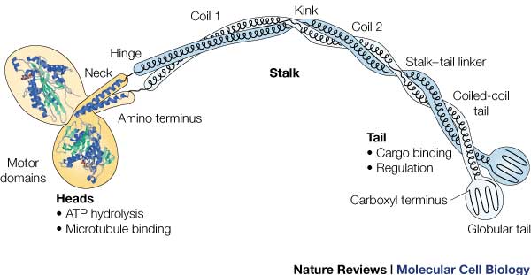
| - | [[Image:131631646354632.jpg]] This is the overall structure of a functional kinesin dimer. | ||
| Line 21: | Line 20: | ||
It is the most conserved region amongst kinesin which consists of the head, neck, and tail. Usually contains eight core β-sheets and six major alpha helixes, most of these secondary structures are in different places in the primary sequence but line up in the tertiary structure. | It is the most conserved region amongst kinesin which consists of the head, neck, and tail. Usually contains eight core β-sheets and six major alpha helixes, most of these secondary structures are in different places in the primary sequence but line up in the tertiary structure. | ||
| - | + | [[Image:Hirokawa.jpg | thumb | Here is a picture of the light chains.]] | |
'''''Light Chain''''' | '''''Light Chain''''' | ||
| - | + | Not technically part of the protein of kinesin or myosin itself, but its presence is necessary for activity. It regulates conformational changes within the protein. | |
| + | [[Image:131631646354632.jpg | This is the overall structure of a functional kinesin dimer.]] | ||
| + | |||
'''Head''' | '''Head''' | ||
Revision as of 00:32, 16 December 2015
Kinesin
| |||||||||||
References
- ↑ Hanson, R. M., Prilusky, J., Renjian, Z., Nakane, T. and Sussman, J. L. (2013), JSmol and the Next-Generation Web-Based Representation of 3D Molecular Structure as Applied to Proteopedia. Isr. J. Chem., 53:207-216. doi:http://dx.doi.org/10.1002/ijch.201300024
- ↑ Herraez A. Biomolecules in the computer: Jmol to the rescue. Biochem Mol Biol Educ. 2006 Jul;34(4):255-61. doi: 10.1002/bmb.2006.494034042644. PMID:21638687 doi:10.1002/bmb.2006.494034042644


