This old version of Proteopedia is provided for student assignments while the new version is undergoing repairs. Content and edits done in this old version of Proteopedia after March 1, 2026 will eventually be lost when it is retired in about June of 2026.
Apply for new accounts at the new Proteopedia. Your logins will work in both the old and new versions.
Diphtheria toxin
From Proteopedia
(Difference between revisions)
| Line 15: | Line 15: | ||
== Structural highlights == | == Structural highlights == | ||
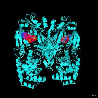
| - | DT is proteolitically cleaved into 2 fragments. Fragment A contains the catalytic domain (C) and fragment B contains the transmembrane (T) and receptor-binding (R) domains. DT active site is located in a cleft in the C domain.<ref>PMID:7833807</ref> The 2 monomers of DT interact by domain swapping to form a compact, globular dimer structure. | + | DT is proteolitically cleaved into 2 fragments. Fragment A contains the <scene name='59/592684/Cv/3'>catalytic domain (C)</scene> and fragment B contains the transmembrane (T) and receptor-binding (R) domains. DT active site is located in a cleft in the C domain.<ref>PMID:7833807</ref> The 2 monomers of DT interact by domain swapping to form a compact, globular dimer structure. |
</StructureSection> | </StructureSection> | ||
Revision as of 15:40, 3 January 2016
| |||||||||||
3D structures of diphtheria toxin
Updated on 03-January-2016
References
- ↑ Pappenheimer AM Jr. Diphtheria toxin. Annu Rev Biochem. 1977;46:69-94. PMID:20040 doi:http://dx.doi.org/10.1146/annurev.bi.46.070177.000441
- ↑ Bennett MJ, Choe S, Eisenberg D. Refined structure of dimeric diphtheria toxin at 2.0 A resolution. Protein Sci. 1994 Sep;3(9):1444-63. PMID:7833807
