We apologize for Proteopedia being slow to respond. For the past two years, a new implementation of Proteopedia has been being built. Soon, it will replace this 18-year old system. All existing content will be moved to the new system at a date that will be announced here.
Sandbox Reserved 1125
From Proteopedia
(Difference between revisions)
| Line 40: | Line 40: | ||
The zinc-binding motif HEXGHXXGXXH presents in the catalytic domain is characteristic for the protease activity of MMP-8. | The zinc-binding motif HEXGHXXGXXH presents in the catalytic domain is characteristic for the protease activity of MMP-8. | ||
===== Zn999 : the catalytic zinc ===== | ===== Zn999 : the catalytic zinc ===== | ||
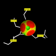
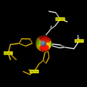
| - | It is involved in the catalytic activity and is situated at the bottom of the active-site. | + | It is involved in the catalytic activity and is situated at the bottom of the active-site. This ion is penta-coordinated with: His197, His201 and His207 of MMP8 and with the carbonyl and the hydroxyl oxygen of the hydroxamic acid moiety of the inhibitor. This discovery has been made thanks to the Pro-Leu-Gly-hydroxylamine inhibitor.<ref>PMID:8137810</ref> On this <scene name='71/719866/Zn999_interactions/2'>link</scene> you can only see the 3 His of MMP8 with the Zn999. The fourth ligand of the catalytic zinc is a water molecule. |
[[Image:ZN pocket interaction.gif | thumb|ZN999 pocket interaction]] | [[Image:ZN pocket interaction.gif | thumb|ZN999 pocket interaction]] | ||
===== Zn998 : the structural zinc ===== | ===== Zn998 : the structural zinc ===== | ||
| Line 49: | Line 49: | ||
<font color='red'>The conserved cysteine present in the cysteine-switch motif (89-96) binds the catalytic zinc ion, thus inhibiting the enzyme. The dissociation of the cysteine from the zinc ion upon the activation-peptide release activates the enzyme.</font> | <font color='red'>The conserved cysteine present in the cysteine-switch motif (89-96) binds the catalytic zinc ion, thus inhibiting the enzyme. The dissociation of the cysteine from the zinc ion upon the activation-peptide release activates the enzyme.</font> | ||
| - | |||
| - | |||
| - | <scene name='71/719866/Catalytic_site/2'>Catalytic site</scene> | ||
=== Hemopexin domain === | === Hemopexin domain === | ||
| Line 69: | Line 66: | ||
| - | Cleavage at Gly775–Ile776 or Leu776 in each alpha-chain of the molecule. <ref>PMID:9094424</ref> The cleavage generates fragments that spontaneously lose their helical conformation, denature to gelatin, and become soluble. The gelatin is then susceptible to attack by gelatinases and other proteases.<ref>[http://www.ebi.ac.uk/interpro/entry/IPR028709 "Neutrophil collagenase"]</ref> | + | Cleavage at Gly775–Ile776 or Leu776 in each alpha-chain of the molecule by the residues of the <scene name='71/719866/catalytic_site/2'>Catalytic site</scene>. <ref>PMID:9094424</ref> The cleavage generates fragments that spontaneously lose their helical conformation, denature to gelatin, and become soluble. The gelatin is then susceptible to attack by gelatinases and other proteases.<ref>[http://www.ebi.ac.uk/interpro/entry/IPR028709 "Neutrophil collagenase"]</ref> |
Revision as of 07:20, 29 January 2016
MMP8
MMP-8, also called, Neutrophil collagenase or Collagenase 2, is a zinc-dependent and calcium-dependent enzyme. It belongs to the matrix metalloproteinase (MMP) family which is involved in the breakdown of extracellular matrix in embryonic development, reproduction, and tissue remodeling, as well as in disease processes, such as arthritis and metastasis. The gene coding this family is localized on the chromosome 11 of Homo sapiens.[1]
| |||||||||||
References
- ↑ "MMP8 matrix metallopeptidase 8 (neutrophil collagenase)"
- ↑ "Metalloendopeptidase activity"
- ↑ Stams T, Spurlino JC, Smith DL, Wahl RC, Ho TF, Qoronfleh MW, Banks TM, Rubin B. Structure of human neutrophil collagenase reveals large S1' specificity pocket. Nat Struct Biol. 1994 Feb;1(2):119-23. PMID:7656015
- ↑ 4.0 4.1 Substrate specificity of MMPs
- ↑ Bode W, Reinemer P, Huber R, Kleine T, Schnierer S, Tschesche H. The X-ray crystal structure of the catalytic domain of human neutrophil collagenase inhibited by a substrate analogue reveals the essentials for catalysis and specificity. EMBO J. 1994 Mar 15;13(6):1263-9. PMID:8137810
- ↑ Hirose T, Patterson C, Pourmotabbed T, Mainardi CL, Hasty KA. Structure-function relationship of human neutrophil collagenase: identification of regions responsible for substrate specificity and general proteinase activity. Proc Natl Acad Sci U S A. 1993 Apr 1;90(7):2569-73. PMID:8464863
- ↑ Knauper V, Osthues A, DeClerck YA, Langley KE, Blaser J, Tschesche H. Fragmentation of human polymorphonuclear-leucocyte collagenase. Biochem J. 1993 May 1;291 ( Pt 3):847-54. PMID:8489511
- ↑ Welgus HG, Jeffrey JJ, Eisen AZ. Human skin fibroblast collagenase. Assessment of activation energy and deuterium isotope effect with collagenous substrates. J Biol Chem. 1981 Sep 25;256(18):9516-21. PMID:6270090
- ↑ Visse R, Nagase H. Matrix metalloproteinases and tissue inhibitors of metalloproteinases: structure, function, and biochemistry. Circ Res. 2003 May 2;92(8):827-39. PMID:12730128 doi:http://dx.doi.org/10.1161/01.RES.0000070112.80711.3D
- ↑ Knauper V, Docherty AJ, Smith B, Tschesche H, Murphy G. Analysis of the contribution of the hinge region of human neutrophil collagenase (HNC, MMP-8) to stability and collagenolytic activity by alanine scanning mutagenesis. FEBS Lett. 1997 Mar 17;405(1):60-4. PMID:9094424
- ↑ "Neutrophil collagenase"
- ↑ "Extra Binding Region Induced by Non-Zinc Chelating Inhibitors into the S1′ Subsite of Matrix Metalloproteinase 8"
- ↑ Balbin M, Fueyo A, Knauper V, Pendas AM, Lopez JM, Jimenez MG, Murphy G, Lopez-Otin C. Collagenase 2 (MMP-8) expression in murine tissue-remodeling processes. Analysis of its potential role in postpartum involution of the uterus. J Biol Chem. 1998 Sep 11;273(37):23959-68. PMID:9727011
RESSOURCE : Image:2oy4 mm1.pdb ( la structure du monomère )


