This old version of Proteopedia is provided for student assignments while the new version is undergoing repairs. Content and edits done in this old version of Proteopedia after March 1, 2026 will eventually be lost when it is retired in about June of 2026.
Apply for new accounts at the new Proteopedia. Your logins will work in both the old and new versions.
Z-DNA model tour
From Proteopedia
(Difference between revisions)
| Line 33: | Line 33: | ||
You can see <scene name='72/725870/Zoom_pairs_only/1'>the same view without the backbone</scene> here.Going 5' to 3', there is good stacking within the GpC dinucleotide, but not between them (CpG). | You can see <scene name='72/725870/Zoom_pairs_only/1'>the same view without the backbone</scene> here.Going 5' to 3', there is good stacking within the GpC dinucleotide, but not between them (CpG). | ||
| - | + | A <scene name='72/725870/Zoom_pair_top/1'>top view</scene> also illustrates the stacking arrangement. You can also see this <scene name='72/725870/Zoom_pairs_only_top/1'>top view of just the bases.</scene>. Note the stacking of red base pairs on each other is much different than the stacking of red on blue. | |
| - | + | ||
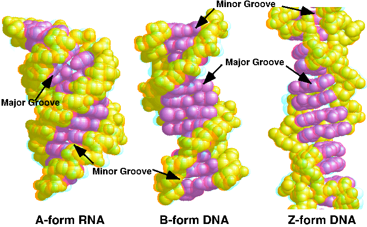
| - | You can compare it with the DNA forms by looking at this [http://proteopedia.org/wiki/images/d/d3/JnABZ3d.gif 3D red-blue stereo picture of A, B, and Z DNA] | + | You can compare it with the other DNA forms by looking at this [http://proteopedia.org/wiki/images/d/d3/JnABZ3d.gif 3D red-blue stereo picture of A, B, and Z DNA] |
</StructureSection> | </StructureSection> | ||
== References == | == References == | ||
Revision as of 20:59, 21 February 2016
Z-form DNA model
| |||||||||||
References
R. E. Dickerson, H. R. Drew, B. N. Conner, R. M. Wing, A. V. Fratini & M. L. Kopka (1982) The anatomy of A-, B-, and Z-DNA. Science 216: 475-485 [1] JSmol in Proteopedia [2] or to the article describing Jmol [3] to the rescue.
