Glucuronidase
From Proteopedia
(Difference between revisions)
| Line 1: | Line 1: | ||
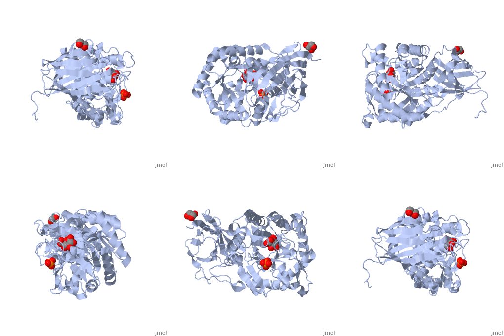
| - | <StructureSection load=' | + | <StructureSection load='3vnz' size='340' side='right' caption='Ribbon diagram of glycosylated human βα-glucuronidase complex with MPD(PDB code [[3hn3]]).' scene=''> |
This tutorial illustrates the quaternary structures of the human and ''E. coli'' β-glucuronidase enzyme. | This tutorial illustrates the quaternary structures of the human and ''E. coli'' β-glucuronidase enzyme. | ||
Revision as of 13:26, 7 March 2016
| |||||||||||
3D Structures of glucuronisidase
Updated on 07-March-2016
References
- ↑ Jain S, Drendel WB, Chen ZW, Mathews FS, Sly WS, Grubb JH. Structure of human beta-glucuronidase reveals candidate lysosomal targeting and active-site motifs. Nat Struct Biol. 1996 Apr;3(4):375-81. PMID:8599764
- ↑ Nagy T, Emami K, Fontes CM, Ferreira LM, Humphry DR, Gilbert HJ. The membrane-bound alpha-glucuronidase from Pseudomonas cellulosa hydrolyzes 4-O-methyl-D-glucuronoxylooligosaccharides but not 4-O-methyl-D-glucuronoxylan. J Bacteriol. 2002 Sep;184(17):4925-9. PMID:12169619
- ↑ doi: https://dx.doi.org/10.2210/pdb3lpf/pdb
- ↑ Sparreboom A, de Jonge MJ, de Bruijn P, Brouwer E, Nooter K, Loos WJ, van Alphen RJ, Mathijssen RH, Stoter G, Verweij J. Irinotecan (CPT-11) metabolism and disposition in cancer patients. Clin Cancer Res. 1998 Nov;4(11):2747-54. PMID:9829738
- ↑ doi: https://dx.doi.org/10.2210/pdb3hn3/pdb

