We apologize for Proteopedia being slow to respond. For the past two years, a new implementation of Proteopedia has been being built. Soon, it will replace this 18-year old system. All existing content will be moved to the new system at a date that will be announced here.
From Proteopedia
(Difference between revisions)
proteopedia linkproteopedia link
|
|
| Line 1: |
Line 1: |
| | ===Monomers Per Turn=== | | ===Monomers Per Turn=== |
| | + | |
| | + | |
| | + | <div class="sketchfab-embed-wrapper"><iframe width="640" height="480" src="https://sketchfab.com/models/f50ee70436064607940be49a356990e6/embed" frameborder="0" allowvr allowfullscreen mozallowfullscreen="true" webkitallowfullscreen="true" onmousewheel=""></iframe> |
| | + | |
| | + | |
| | | | |
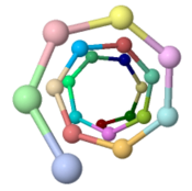
| | The ARC-1 model has 6.4 monomer chains per turn (56.0 degrees rotation between monomers). This is more chains/turn than some previous type IV pilus models. | | The ARC-1 model has 6.4 monomer chains per turn (56.0 degrees rotation between monomers). This is more chains/turn than some previous type IV pilus models. |
Revision as of 21:15, 4 March 2017
Monomers Per Turn
<iframe width="640" height="480" src="
https://sketchfab.com/models/f50ee70436064607940be49a356990e6/embed" frameborder="0" allowvr allowfullscreen mozallowfullscreen="true" webkitallowfullscreen="true" onmousewheel=""></iframe>
The ARC-1 model has 6.4 monomer chains per turn (56.0 degrees rotation between monomers). This is more chains/turn than some previous type IV pilus models.
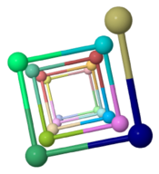
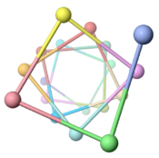
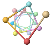
To visualize chains/turn, we show (alpha carbon of Phe51). Then these chain-marking-atoms are , and the resulting helix is viewed from one end. When counting the chains/turn, bear in mind that the first and last (to complete one turn) count as 1/2 chain each.
|
Pilus
|
Chains/Turn (Angle)
|
Image
|
|
Geobacter sulfurreducens (type IVa, 61 amino acids, theoretical model, 2016)[1]
|
6.4 (56.0°)
|

|
|
Pseudomonas aeruginosa (type IVa, 150 amino acids, fiber diffraction, 2004)[2]
|
4.0 (90°)
|

|
|
Vibrio cholerae (type IVb, 198 amino acids, cryo-EM, 2012)[3]
|
3.7 (96.8°)
|

|
|
Neisseria gonorrhoeae (type IVa, 165 amino acids, cryo-EM, 2006)[4]
|
3.6 (100.8°)
|

|
Notes & References
- ↑ Cite error: Invalid
<ref> tag;
no text was provided for refs named xiao1
- ↑ Craig L, Pique ME, Tainer JA. Type IV pilus structure and bacterial pathogenicity. Nat Rev Microbiol. 2004 May;2(5):363-78. PMID:15100690 doi:http://dx.doi.org/10.1038/nrmicro885
- ↑ Li J, Egelman EH, Craig L. Structure of the Vibrio cholerae Type IVb Pilus and stability comparison with the Neisseria gonorrhoeae type IVa pilus. J Mol Biol. 2012 Apr 20;418(1-2):47-64. doi: 10.1016/j.jmb.2012.02.017. Epub 2012, Feb 21. PMID:22361030 doi:http://dx.doi.org/10.1016/j.jmb.2012.02.017
- ↑ Craig L, Volkmann N, Arvai AS, Pique ME, Yeager M, Egelman EH, Tainer JA. Type IV pilus structure by cryo-electron microscopy and crystallography: implications for pilus assembly and functions. Mol Cell. 2006 Sep 1;23(5):651-62. PMID:16949362 doi:10.1016/j.molcel.2006.07.004


