We apologize for Proteopedia being slow to respond. For the past two years, a new implementation of Proteopedia has been being built. Soon, it will replace this 18-year old system. All existing content will be moved to the new system at a date that will be announced here.
Phosphoglycerate Mutase
From Proteopedia
(Difference between revisions)
| Line 1: | Line 1: | ||
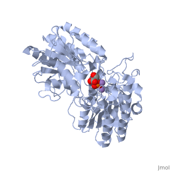
| - | + | <StructureSection load='1eqj' size='400' side='right' caption='Phosphoglycerate mutase complex with phosphoglyceric acid and Mn+2 ion (purple) [[1eqj]]' scene='' pspeed='8'> | |
== Background == | == Background == | ||
| Line 19: | Line 19: | ||
It is important to note that the phosphate group that is placed on C2 is not the same phosphate group that was initially on C3. | It is important to note that the phosphate group that is placed on C2 is not the same phosphate group that was initially on C3. | ||
| - | In order to understand how PGM catalyzes this reaction, an explanation of its active site is imperative. The most important residues in this enzyme include <scene name='Christopher_Vachon_Sandbox/His_8_good/2'>His 8 and 179</scene> with imidazole groups which are in close proximity to carbons 2 and 3 in the substrate. His-8 is phosphorylated during during catalysis, and it is likely that His-179 acts as the proton donor/acceptor <ref>Rose, Z.B. (1980) Adv. Enzymol. Relat. Areas Mol. Biol. 51, | + | In order to understand how PGM catalyzes this reaction, an explanation of its active site is imperative. The most important residues in this enzyme include <scene name='Christopher_Vachon_Sandbox/His_8_good/2'>His 8 and 179</scene> with imidazole groups which are in close proximity to carbons 2 and 3 in the substrate. His-8 is phosphorylated during during catalysis, and it is likely that His-179 acts as the proton donor/acceptor <ref>Rose, Z.B. (1980) Adv. Enzymol. Relat. Areas Mol. Biol. 51, 211-253</ref>. Based on crystallography experiments, the active site where these histidine residues reside lies at the bottom of a deep groove in each subunit. <ref name="winn" /> The sites in each subunit, whether the enzyme is a homodimer or homotetramer, are well separated. The active enzyme contains a phosphoryl group attached to His 8. This phosphoryl group is what is transferred to C2 of the substrate, resulting in an intermediate 2,3-bisphosphoglycerate-enzyme complex. Thus there is a <scene name='Christopher_Vachon_Sandbox/Good_active_site_scene/6'>covalently attached phosphate</scene> in the active monomer. <ref name="voet" /> The phosphate group on C3 of the substrate is then transferred back onto His 8, thus regenerating the active form of the enzyme. |
In addition to the importance of the two histidine residues in the active site, the amino acids that line the <scene name='Christopher_Vachon_Sandbox/Good_active_site_scene/5'>active site</scene> are also functionally important. These residues include H179, H8, E15, S11, T20, R59, and E86.<ref name="voet" /> Several positively charged residues line the active site pocket. These residues usually tend to be <scene name='Christopher_Vachon_Sandbox/Arginine_residues/1'>arginine residues</scene>, which are important for the optimal activity of the enzyme. <ref name="winn" /> This structure is logical for its function because the enzyme binds a negatively charged substrate, thus a positively charged groove fosters tight binding with a negative substrate. The third and final important aspect of the active site is the presence of <scene name='Christopher_Vachon_Sandbox/Glutamate_residues_2/1'>glutamate residues 15 and 86</scene>.<ref name="winn" /> It is suggested that the carboxyl groups of these amino acid residues act as proton-withdrawing groups as they flank both sides of the substrate. | In addition to the importance of the two histidine residues in the active site, the amino acids that line the <scene name='Christopher_Vachon_Sandbox/Good_active_site_scene/5'>active site</scene> are also functionally important. These residues include H179, H8, E15, S11, T20, R59, and E86.<ref name="voet" /> Several positively charged residues line the active site pocket. These residues usually tend to be <scene name='Christopher_Vachon_Sandbox/Arginine_residues/1'>arginine residues</scene>, which are important for the optimal activity of the enzyme. <ref name="winn" /> This structure is logical for its function because the enzyme binds a negatively charged substrate, thus a positively charged groove fosters tight binding with a negative substrate. The third and final important aspect of the active site is the presence of <scene name='Christopher_Vachon_Sandbox/Glutamate_residues_2/1'>glutamate residues 15 and 86</scene>.<ref name="winn" /> It is suggested that the carboxyl groups of these amino acid residues act as proton-withdrawing groups as they flank both sides of the substrate. | ||
| Line 31: | Line 31: | ||
== Phosphoglycerate Mutase Deficiency == | == Phosphoglycerate Mutase Deficiency == | ||
When phosphoglycerate mutase has a genetic defect, it results in a muscle disease that interferes with the processing of carbohydrates. The onset can occur anywhere from childhood to adulthood. The inheritance pattern is autosomal recessive. <ref>http://www.mda.org/disease/pgam.html</ref> Phosphoglycerate mutase deficicncy patients may experience CNS symptoms such as mental retardation and seizures. Certain individuals may experience a purely myopathic syndrome with progressive proximal muscle weakness and incidents of myoglobinuria, exercise intolerance, may become easy fatigued with cramps and urine discoloration. Diagnosing this deficiency can be done with Laboratory tests that demonstrate and increased serum CK level. Or there are diagnostic tests available that test for the absence of the enzyme. Also, muscle pathology of this deficiency shows subsarcolemmal glycogen ± tubular combinations. <ref>http://disability.ucdavis.edu/disease_deatails.php?id=45</ref> | When phosphoglycerate mutase has a genetic defect, it results in a muscle disease that interferes with the processing of carbohydrates. The onset can occur anywhere from childhood to adulthood. The inheritance pattern is autosomal recessive. <ref>http://www.mda.org/disease/pgam.html</ref> Phosphoglycerate mutase deficicncy patients may experience CNS symptoms such as mental retardation and seizures. Certain individuals may experience a purely myopathic syndrome with progressive proximal muscle weakness and incidents of myoglobinuria, exercise intolerance, may become easy fatigued with cramps and urine discoloration. Diagnosing this deficiency can be done with Laboratory tests that demonstrate and increased serum CK level. Or there are diagnostic tests available that test for the absence of the enzyme. Also, muscle pathology of this deficiency shows subsarcolemmal glycogen ± tubular combinations. <ref>http://disability.ucdavis.edu/disease_deatails.php?id=45</ref> | ||
| - | + | </StructureSection> | |
==3D structures of phosphoglycerate mutase== | ==3D structures of phosphoglycerate mutase== | ||
Revision as of 13:29, 31 August 2017
| |||||||||||


3D structures of phosphoglycerate mutase
Updated on 31-August-2017
Additional Resources
For additional information, please see: Carbohydrate Metabolism
References
- ↑ Crowhurst GS, Dalby AR, Isupov MN, Campbell JW, Littlechild JA. Structure of a phosphoglycerate mutase:3-phosphoglyceric acid complex at 1.7 A. Acta Crystallogr D Biol Crystallogr. 1999 Nov;55(Pt 11):1822-6. PMID:10531478
- ↑ http://disability.ucdavis.edu/disease_deatails.php?id=45
- ↑ 3.0 3.1 3.2 3.3 3.4 3.5 S., Winn I., Fothergill A. L., Harkins N. R., and Watson C. H. "Structure and Activity of Phosphoglycerate Mutase." Sciences 293.1063 (1981): 121-30. Print.
- ↑ "Phosphoglycerate mutase -." Wikipedia, the free encyclopedia. Web. 27 Feb. 2010. <http://en.wikipedia.org/wiki/Phosphoglycerate_mutase>.
- ↑ 5.0 5.1 5.2 Voet, Donald, Judith G. Voet, and Charlotte W. Pratt. Fundamentals of Biochemistry Life at the Molecular Level. New York: John Wiley & Sons, 2008. Print.
- ↑ Rose, Z.B. (1980) Adv. Enzymol. Relat. Areas Mol. Biol. 51, 211-253
- ↑ Rigden, D. J.; Walter, R. A.; Phillips, S. E. V.; Fothergill-Gilmore, L. A.Polyanionic inhibitors of phosphoglycerate mutase: combined structural and biochemical analysis J. Mol. Biol. 1999, 289, 691– 699
- ↑ McAleese, S.M., Fothergill-Gilmore, L.A.&Dixon, H.B.F. (1985) Biochem. J. 230, 535-542
- ↑ http://www.mda.org/disease/pgam.html
- ↑ http://disability.ucdavis.edu/disease_deatails.php?id=45
Proteopedia Page Contributors and Editors (what is this?)
Michal Harel, Alexander Berchansky, Robert Trahin, Xuan Loi, David Canner, Christopher Vachon, Allie Paton
