We apologize for Proteopedia being slow to respond. For the past two years, a new implementation of Proteopedia has been being built. Soon, it will replace this 18-year old system. All existing content will be moved to the new system at a date that will be announced here.
Sandbox Reserved 1160
From Proteopedia
(Difference between revisions)
| Line 16: | Line 16: | ||
When mGlu5 is in the active conformation, signaling begins with glutamate binding to the Venus flytrap domain. The signal is transduced across the cystine-rich domain to the TMD<ref name="Niswender" />. Next, the dimerization of the TMD occurs. This activates the Gq/11 pathway, which activates phospholipase Cβ<ref name="Niswender" />. The active [http://www.proteopedia.org/wiki/index.php/2zkm phospholipase Cβ] hydrolyzes phosphotinositides and generates [https://pubchem.ncbi.nlm.nih.gov/compound/439456#section=Top inositol 1,4,5-trisphosphate] and [http://www.sivabio.50webs.com/ip3.htm diacyl-glycerol]<ref name="Woodcock" />. This results in calcium mobilization and activation of protein kinase C (PKC)<ref name="Niswender" />. Calcium is a neurotransmitter, and relatively low concentrations of calcium can cause a large response across the neuronal synapse<ref name="Niswender" />. In addition to calcium stimulating an exitory response in nerve cells, PKC can be activated for regulatory purposes with the influx of calcium. A serine on PKC can become phosphorylated which leaves it unable to bind to G beta-gamma (Gßγ) protein complex<ref name="Niswender" />. Unbound Gßγ protein can then inhibit voltage-sensitive calcium channels to reduce calcium influx and provide feedback inhibition to the glutamate signaling pathway. <ref name="Niswender" />. | When mGlu5 is in the active conformation, signaling begins with glutamate binding to the Venus flytrap domain. The signal is transduced across the cystine-rich domain to the TMD<ref name="Niswender" />. Next, the dimerization of the TMD occurs. This activates the Gq/11 pathway, which activates phospholipase Cβ<ref name="Niswender" />. The active [http://www.proteopedia.org/wiki/index.php/2zkm phospholipase Cβ] hydrolyzes phosphotinositides and generates [https://pubchem.ncbi.nlm.nih.gov/compound/439456#section=Top inositol 1,4,5-trisphosphate] and [http://www.sivabio.50webs.com/ip3.htm diacyl-glycerol]<ref name="Woodcock" />. This results in calcium mobilization and activation of protein kinase C (PKC)<ref name="Niswender" />. Calcium is a neurotransmitter, and relatively low concentrations of calcium can cause a large response across the neuronal synapse<ref name="Niswender" />. In addition to calcium stimulating an exitory response in nerve cells, PKC can be activated for regulatory purposes with the influx of calcium. A serine on PKC can become phosphorylated which leaves it unable to bind to G beta-gamma (Gßγ) protein complex<ref name="Niswender" />. Unbound Gßγ protein can then inhibit voltage-sensitive calcium channels to reduce calcium influx and provide feedback inhibition to the glutamate signaling pathway. <ref name="Niswender" />. | ||
=== Extracellular Domain === | === Extracellular Domain === | ||
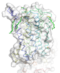
| - | + | The extracellular domain of mGlu5 contains several key extracellular loops that will help modulate ligand binding. <scene name='72/721532/Ecl_trail_7/2'>Extracellular Loops</scene> are shown here with extracellular loops (ECLs) <font color='purple'><b>1</b></font>, <font color='purple'><b>2</b></font>, and <font color='purple'><b>3</b></font> highlighted in <font color='purple'><b>purple</b></font>. Additionally in the ECL domain, a <scene name='72/721531/Ecl_trail_1/5'>disulfide bond</scene> is attached to both Helix 3 and the amino acid chain between Helix 5 and the N-terminus. <font color='teal'><b>Helix 3</b></font> and <font color='red'><b>Helix 5</b></font> are colored in <font color='teal'><b>teal</b></font> and <font color='red'><b>red</b></font> respectively. <font color='blue'><b>N-terminus</b></font> is represented in <font color='blue'><b>blue</b></font>. The <span style="color:yellow;background-color:black;font-weight:bold;">disulfide bond</span> is highlighted in <span style="color:yellow;background-color:black;font-weight:bold;">yellow</span>, and it is conserved in all classes of mGlu5 TMD<ref name="Wu" />. The disulfide bond is critical in maintaining the position of ECL 2 <ref name="Dore" />. ECLs and the helices are also factors that dictate how mavolgurent fits in the binding pocket <ref name="Dore" />. The position of these ECLs can change the effective size of the binding pocket through loop positioning<ref name="Dore" />. | |
=== Binding Pocket === | === Binding Pocket === | ||
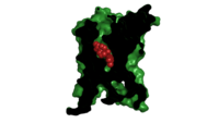
[[Image: Organic with clipped surface.png|200 px|left|thumb|'''Figure 2.''' Mavolugarent in its binding pocket of the 7TM region of mGLu5 Class C receptor. The binding pocket's surface is clipped in black with the substrate, mavolugarent, in red. The rest of the protein is colored in green. The binding pocket is present in the 7 Helix Transmembrane Domain that would be present in the phosolipid bilayer appearing as an intergral protein. The presence of mavolugarent inhibits the function of the metabotropic glutamate receptor.]] | [[Image: Organic with clipped surface.png|200 px|left|thumb|'''Figure 2.''' Mavolugarent in its binding pocket of the 7TM region of mGLu5 Class C receptor. The binding pocket's surface is clipped in black with the substrate, mavolugarent, in red. The rest of the protein is colored in green. The binding pocket is present in the 7 Helix Transmembrane Domain that would be present in the phosolipid bilayer appearing as an intergral protein. The presence of mavolugarent inhibits the function of the metabotropic glutamate receptor.]] | ||
| - | The binding pocket, not to be confused with the glutamate binding site, represents an useful source of regulatory control. The binding pocket is only accessible by a relatively narrow (7 Å) <scene name='72/721531/Bindingsite/1'>entrance</scene>. This small entrance severely restricts the access of both positive and negative allosteric regulators. One such regulator, <scene name='72/721531/Mavo/2'>mavoglurant</scene>, is a relatively small molecule that can travel through the entrance and fit between the helices of the mGlu5 receptor. Mavoglurant acts as a negative allosteric modulator of the mGlu5 receptor. Mavoglurant is a relatively hydrophobic molecule, which compliments the largely hydrophobic nature of the interior of mGlu5. The carbamate tail and hydroxyl group of mavoglurant will also interact with several key amino acids in the binding site of mGlu5. | + | The binding pocket, not to be confused with the glutamate binding site, represents an useful source of regulatory control. The binding pocket is only accessible by a relatively narrow (7 Å) <scene name='72/721531/Bindingsite/1'>entrance</scene><ref name="Dore" />. This small entrance severely restricts the access of both positive and negative allosteric regulators. One such regulator, <scene name='72/721531/Mavo/2'>mavoglurant</scene>, is a relatively small molecule that can travel through the entrance and fit between the helices of the mGlu5 receptor<ref name="Dore" />. Mavoglurant acts as a negative allosteric modulator of the mGlu5 receptor<ref name="Dore" />. Mavoglurant is a relatively hydrophobic molecule, which compliments the largely hydrophobic nature of the interior of mGlu5<ref name="Dore" />. The carbamate tail and hydroxyl group of mavoglurant will also interact with several key amino acids in the binding site of mGlu5<ref name="Dore" />. |
Important Amino Acids<ref name="Dore" />: | Important Amino Acids<ref name="Dore" />: | ||
| Line 27: | Line 27: | ||
*A <scene name='72/721531/Protien_bindbottom/2'>water molecule</scene> inside of the binding pocket helps stabilize the inactive state. | *A <scene name='72/721531/Protien_bindbottom/2'>water molecule</scene> inside of the binding pocket helps stabilize the inactive state. | ||
| - | Upon mavoglurant’s diffusion into the mGlu5 receptor binding pocket, the ordered water cage found in the center of mGlu5 is displaced. Favorable interactions between the hydrophobic regions of the binding pocket and the bicyclic ring and the 3-methylphenyl ring of mavoglurant help increase the strength and energetic favorability of mavoglurant binding via the hydrophobic effect. In the binding pocket mavoglurant can hydrogen bond with Asp 747 and that hydrogen bond will be further stabilized by an extended hydrogen bond network with Gly 652. The hydroxyl group of mavoglurant will also form another hydrogen bond with Ser 805 and Ser 809. Multiple hydrogen bonds make the binding of mavoglurant to the mGlu5 receptor favorable and specific. Once bound to mavoglurant, transmembrane helix 7 undergoes a conformational change<ref name="Dore" />. The shifting of TM7 will lead to a more global conformational change, which inactivates the receptor<ref name="Dore" />. Variation can be seen in positioning of alpha helices across receptor class. Class C receptors have | + | Upon mavoglurant’s diffusion into the mGlu5 receptor binding pocket, the ordered water cage found in the center of mGlu5 is displaced<ref name="Dore" />. Favorable interactions between the hydrophobic regions of the binding pocket and the bicyclic ring and the 3-methylphenyl ring of mavoglurant help increase the strength and energetic favorability of mavoglurant binding via the hydrophobic effect. In the binding pocket mavoglurant can hydrogen bond with Asp 747 and that hydrogen bond will be further stabilized by an extended hydrogen bond network with Gly 652<ref name="Dore" />. The hydroxyl group of mavoglurant will also form another hydrogen bond with Ser 805 and Ser 809<ref name="Dore" />. Multiple hydrogen bonds make the binding of mavoglurant to the mGlu5 receptor favorable and specific<ref name="Wu" />. Once bound to mavoglurant, transmembrane helix 7 undergoes a conformational change<ref name="Dore" />. The shifting of TM7 will lead to a more global conformational change, which inactivates the receptor<ref name="Dore" />. Variation can be seen in positioning of alpha helices 5 and 7 across receptor class. Class C receptors have less space for mavoglurant to enter as compared to Class A and F receptors<ref name="Wu" />. Increased specificity and stronger binding affinity could be a result of the more narrow structure of mGlu5. |
=== Ionic Locks === | === Ionic Locks === | ||
Revision as of 03:40, 19 April 2016
Human metabotropic glutamate receptor 5 transmembrane domain
| |||||||||||


