P53
From Proteopedia
(Difference between revisions)
| Line 1: | Line 1: | ||
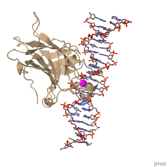
<StructureSection load='1tup' size='350' side='right' scene='26/26327/Initl/2' caption='Human p53 DNA-binding domain complex with DNA and Zn+2 ions (grey) (PDB code [[1tup]])'> | <StructureSection load='1tup' size='350' side='right' scene='26/26327/Initl/2' caption='Human p53 DNA-binding domain complex with DNA and Zn+2 ions (grey) (PDB code [[1tup]])'> | ||
[[Image:P53_DNA.png|left|150px]]<br /> | [[Image:P53_DNA.png|left|150px]]<br /> | ||
| - | + | ||
| - | + | ||
=='''p53 Tumor Suppressor Protein (PDB code [[1tup]])'''== | =='''p53 Tumor Suppressor Protein (PDB code [[1tup]])'''== | ||
| - | ---- | ||
'''p53''' is a tumor suppressor. In the absence of cellular stress, p53 does not exert effect on cell fate, but under stress, p53 becomes activated and causes phenotypic changes in cells like senescence and apoptosis<ref>PMID:12719720</ref>. Many human cancer cells carry mutated p53<ref>PMID:20182618</ref>. The name '''p53''' refers to its apparent molecular mass. It runs as a 53 kDa molecule on SDS-PAGE. But based on calculations from its amino acid residues, p53's mass is actually 43.7 kDa. This difference may be due to the high number of proline residues in the protein, resulting in its migrating slowly on SDS-PAGE. | '''p53''' is a tumor suppressor. In the absence of cellular stress, p53 does not exert effect on cell fate, but under stress, p53 becomes activated and causes phenotypic changes in cells like senescence and apoptosis<ref>PMID:12719720</ref>. Many human cancer cells carry mutated p53<ref>PMID:20182618</ref>. The name '''p53''' refers to its apparent molecular mass. It runs as a 53 kDa molecule on SDS-PAGE. But based on calculations from its amino acid residues, p53's mass is actually 43.7 kDa. This difference may be due to the high number of proline residues in the protein, resulting in its migrating slowly on SDS-PAGE. | ||
Revision as of 10:42, 14 January 2017
| |||||||||||


3D structures of p53 (Updated on 14-January-2017)
Additional Resources
References
- ↑ Oren M. Decision making by p53: life, death and cancer. Cell Death Differ. 2003 Apr;10(4):431-42. PMID:12719720 doi:http://dx.doi.org/10.1038/sj.cdd.4401183
- ↑ Oren M, Rotter V. Mutant p53 gain-of-function in cancer. Cold Spring Harb Perspect Biol. 2010 Feb;2(2):a001107. doi:, 10.1101/cshperspect.a001107. PMID:20182618 doi:http://dx.doi.org/10.1101/cshperspect.a001107
Proteopedia Page Contributors and Editors (what is this?)
Joel L. Sussman, Michal Harel, Eran Hodis, Mary Ball, Alexander Berchansky, David Canner
