Overview
PsaA protein
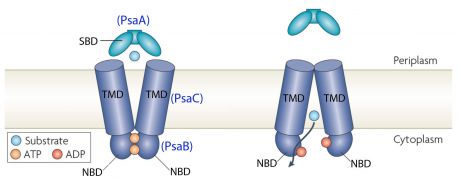
PsaA (Pneumococcal surface antigen A) is a multi-functional lipoprotein detected on all known serotypes of Streptococcus pneumoniae. This lipoprotein is part of the ABC-type (ATP binding complex) transport protein complex that transports Mn2+. PsaA is also an adhesion factor that plays a major role in pneumococcal attachment to the host cell and virulence. PsaA is hidden beneath the cell wall. PsaA protein is involved in in colonization of the nasopharyngeal mucosal.
This transporter is composed of the products of three genes, psaB (ATP-binding protein), psaC (integral membrane protein), and psaA (solute-binding lipoprotein), which are organized in an operon with a gene encoding PsaD, a thiol peroxidase [1].
Between the first to the 24th amino acid namely the red motif there is the signal peptide and the second one is the Pfam motif[3].
Zinc in excess has significant toxicity to bacteria because it is an important innate defence mechanism. There are many Zinc in human body. Manganese is important for the virulence, growth and proliferation of Streptococcus pneumoniae. Zinc could compete for Manganese binding. However Manganese has more affinity for PsaA than Zinc but Zinc is not transported by the ABC-transporter. Zinc competition reduces intracellular Manganse resulting in up-regulation of PsaBCA expression. [4]
Streptococcus pneumoniae
Streptococcus pneumoniae is a Gram positive cocci (with a diameter from 0.5 to 1 μm) and a member of the genus Streptococcus. It can live under aerobic or anaerobic conditions. It resides in the nasopharynx of healthy carriers. However, the bacterium may become pathogenic in elderly and immunocompromised adults and children. Then it can spread to other locations and cause disease. The genome of S. pneumoniae is a closed, circular DNA structure that contains between 2.0 and 2.1 million base pairs.[6]
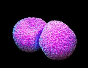
Streptococcus pneumoniae [7]
Structure
The protein PsaA has a molecular weight of 34.538 kDa with 309 residues[8]. The overall size of the protein approximated from its crystal structure is 40 by 40 by 70 Å[9].
As a member of the Lipoprotein receptor-associated antigen I (LraI) family, the PsaA molecule contains four distinct regions. An N-terminal leader sequence of 20 amino acids holds an LxACy consensus sequence that is recognized and cleaved by signal peptidase II [10]. A lipid moiety (diacylglycerol [11]) is added to the cysteine residue and mediates the anchorage of the protein to the cytoplasmic membrane. Apart from this leader sequence, the rest of the protein consists of two twofold-pseudosymmetrical (β/α)4 sandwich domains, of which the β-strands of each domain form parallel β-sheets [12]. In total the two domains form two lobes connected via an α-helical linker which constitutes the solute-binding site [13].
| 
|
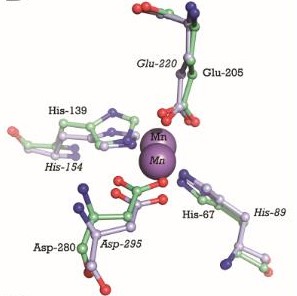
This image shows the metal binding site in more detail, with the MntC residues and Manganese. Manganse ions are shown as purple spheres. We can see that Manganese interacts with Histine residues, Aspartique acid residues and Glutamique acid residues.
The metal binding site is formed by the sidechains of residues His67, His139, Glu205, and Asp280. This site has tetrahedral coordination geometry. The amino acids His67 andHis139 interact with the metal via Nε2 nitrogen atoms. However, the carboxylate sidechains of Glu205 and Asp280 interact with the metal via their Oε1 and Oδ2 atoms. The atomic distance between His67 Nε2 and the metal is 1.99 Ắ while it is 2,01 Ắ for His 139 Nε2. It is 2,04 Ắ for Glu205 Oε1 and 2,02 Ắ for Asp280 Oδ2. The Oδ1 atom of Asp137 can form a hydrogen bond with Nδ1 of His139. Oδ1 atom of Asp65 can also form a hydrogen bond with His67 N. [14]
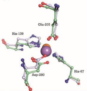
This image shows the metal binding site in more detail, with MntC residues and Managanese and Zinc. Manganese ion is shown as a purple sphere and Zinc ion is shown as an orange sphere. Cadmium uptake reduces the millimolar cellular accumulation of manganese and zinc, and thereby increases sensitivity to oxidative stress [15].
3D structure of PsaA with 2-AMINO-2-HYDROXYMETHYL-PROPANE-1,3-DIOL (tris) and cadmium[16]
Protein-protein interaction
Function
Mn2+ is also required for the activity of CpsB, a tyrosine phosphatase involved in the regulation of capsule production. And in some streptococcal species, lectin-mediated adherence requires Mn2+. Finally, mutations in the psa operon result in an almost complete attenuation of virulence for all tested models of animal infection.
Disease
Meningitis
Otitis
PsaA has been recognized to be involved in the adherence and virulence mechanisms of otitis media. Streptococcus pneumoniae is one of the main agents causing bacterial acute otitis media, directly or as complication of a viral upper respiratory tract infection. This disease is a highly prevalent pediatric disease worldwide. Hearing loss is a common problem associated with this disease. [17]
Pneumonia
PsaA protein is an adhesin which is involved in colonization of the nasopharyngeal mucosal. Moreover, alveolar pneumonia is caused by the spread of Streptococcus pneumoniae from nasopharynx. Therefore PsaA protein is involved in infection of pulmonary parenchyma by Streptococcus pneumoniae.
Sometimes, Streptococcus pneumoniae pass into the blood and causes a bacteremia besides pneumonia.
Application in Biotechnology
PsaA is being actively evaluated as a component of a vaccin in formulations composed of pneumococcal common proteins. PsaA is a component of a vaccin because this protein is immunogenic and stimulates an increase in antibody production when the nasopharynx is naturally colonized. PsaA has been expressed as an E.coli recombinant protein, purified, and evaluated in a phase one clinical trial.
Progress in vaccine development is most advanced for Streptococcus pneumoniae. Indeed, there is a seven-valent capsular-conjugate vaccine, PREVNAR® but it is rather non efficient for otitis media.[18]
References
- ↑ Moore RC. Investigational drug information is available to the pharmacist. Am J Hosp Pharm. 1979 Nov;36(11):1480, 1484. PMID:517531
- ↑ McAllister LJ, Tseng HJ, Ogunniyi AD, Jennings MP, McEwan AG, Paton JC. Molecular analysis of the psa permease complex of Streptococcus pneumoniae. Mol Microbiol. 2004 Aug;53(3):889-901. PMID:15255900 doi:http://dx.doi.org/10.1111/j.1365-2958.2004.04164.x
- ↑ http://string-db.org/cgi/network.pl?taskId=EA2jsQm5Sc5A
- ↑ McDevitt CA, Ogunniyi AD, Valkov E, Lawrence MC, Kobe B, McEwan AG, Paton JC. A molecular mechanism for bacterial susceptibility to zinc. PLoS Pathog. 2011 Nov;7(11):e1002357. Epub 2011 Nov 3. PMID:22072971 doi:10.1371/journal.ppat.1002357
- ↑ http://www.latrobe.edu.au/biochemistry-and-genetics/research/maher/psabca-manganese-uptake-by-streptococcus-pneumoniae
- ↑ Bierman EL, Stein O, Stein Y. Lipoprotein uptake and metabolism by rat aortic smooth muscle cells in tissue culture. Circ Res. 1974 Jul;35(1):136-50. PMID:4366526
- ↑ http://www.sciencephoto.com/images/download_lo_res.html?id=662360183
- ↑ https://www.mybiosource.com/prods/Recombinant-Protein/Manganese-ABC-transporter-substrate-binding-lipoprotein-psaA/psaA
- ↑ Yu Y, Chang, Xu H, Zhang X, Pan L, Xu C, Huang B, Zhou H, Li J, Guo J, Liu C. The virulence of Streptococcus pneumoniae partially depends on dprA. Braz J Microbiol. 2016 Dec 6. pii: S1517-8382(16)30151-4. doi:, 10.1016/j.bjm.2016.10.019. PMID:28011228 doi:http://dx.doi.org/10.1016/j.bjm.2016.10.019
- ↑ Yu Y, Chang, Xu H, Zhang X, Pan L, Xu C, Huang B, Zhou H, Li J, Guo J, Liu C. The virulence of Streptococcus pneumoniae partially depends on dprA. Braz J Microbiol. 2016 Dec 6. pii: S1517-8382(16)30151-4. doi:, 10.1016/j.bjm.2016.10.019. PMID:28011228 doi:http://dx.doi.org/10.1016/j.bjm.2016.10.019
- ↑ PMID:PMC99024
- ↑ PMID:PMC99024
- ↑ Yu Y, Chang, Xu H, Zhang X, Pan L, Xu C, Huang B, Zhou H, Li J, Guo J, Liu C. The virulence of Streptococcus pneumoniae partially depends on dprA. Braz J Microbiol. 2016 Dec 6. pii: S1517-8382(16)30151-4. doi:, 10.1016/j.bjm.2016.10.019. PMID:28011228 doi:http://dx.doi.org/10.1016/j.bjm.2016.10.019
- ↑ Lawrence MC, Pilling PA, Epa VC, Berry AM, Ogunniyi AD, Paton JC. The crystal structure of pneumococcal surface antigen PsaA reveals a metal-binding site and a novel structure for a putative ABC-type binding protein. Structure. 1998 Dec 15;6(12):1553-61. PMID:9862808
- ↑ http://www.nature.com/articles/ncomms7418
- ↑ http://www.ebi.ac.uk/pdbe/entry/pdb/4UTO
- ↑ https://www.google.com/patents/US20130078254
- ↑ Wald ER, Mason EO Jr, Bradley JS, Barson WJ, Kaplan SL. Acute otitis media caused by Streptococcus pneumoniae in children's hospitals between 1994 and 1997. Pediatr Infect Dis J. 2001 Jan;20(1):34-9. PMID:11176564


