Carboxypeptidase A
From Proteopedia
(Difference between revisions)
| Line 25: | Line 25: | ||
==Important Tyr248 Residue== | ==Important Tyr248 Residue== | ||
| - | [http://www.rcsb.org/pdb/explore/explore.do?structureId= | + | CPA from ''B. taurus'' has been co-crystallized with two Zn<sup>2+</sup> ions (Figure 1). This structure has been deposited in the PDB database under the label [http://www.rcsb.org/pdb/explore/explore.do?structureId=1cpx 1CPX], which is a β-form of CPA. 1CPX has an interesting distinction from some other crystallized CPA proteins in that its crystallographic data has revealed a different conformation of the Tyr248 residue [[Image:1CPX-Tyr248.png|thumb|Figure 2: Important Tyr248 residue of 1CPX is pointing toward the hydrophobic binding pocket.]] <ref name="CPA1">Bukrinsky JT, Bjerrum MJ, Kadziola A. 1998. Native carboxypeptidase A in a new crystal environment reveals a different conformation of the important tyrosine 248. ''Biochemistry''. 37(47):16555-16564. [http://pubs.acs.org/doi/abs/10.1021/bi981678i DOI: 10.1021/bi981678i]</ref>. Previous literature has suggested that the conserved Tyrosine residue among CPA proteins has been involved in an [http://en.wikipedia.org/wiki/Enzyme_catalysis#Induced_fit induced fit mechanism] because Tyr248 typically has been found <scene name='69/694222/Tyr248_5cpa/1'>pointing outward</scene> toward solution when a substrate is not bound in the active site.<ref name="CPA1">Bukrinsky JT, Bjerrum MJ, Kadziola A. 1998. Native carboxypeptidase A in a new crystal environment reveals a different conformation of the important tyrosine 248. ''Biochemistry''. 37(47):16555-16564. [http://pubs.acs.org/doi/abs/10.1021/bi981678i DOI: 10.1021/bi981678i]</ref> However, crystallographic data for 1CPX shows Tyr248 <scene name='69/694222/Tyr248_1cpx/1'>pointing toward the active site</scene> without a polypeptide substrate bound. Therefore, this conflicts with the previously proposed induced fit mechanism for CPA proteins and suggests that Tyr248 is liganded to the catalytic Zn<sup>2+</sup> ion in 1CPX.<ref name="CPA1">Bukrinsky JT, Bjerrum MJ, Kadziola A. 1998. Native carboxypeptidase A in a new crystal environment reveals a different conformation of the important tyrosine 248. ''Biochemistry''. 37(47):16555-16564. [http://pubs.acs.org/doi/abs/10.1021/bi981678i DOI: 10.1021/bi981678i]</ref> Moreover, this data supports that this is the <scene name='69/694222/Tyr248_1cpx/1'>native conformation of Tyr248 in solution</scene> because none of the residues in the protein’s active site interact in crystallographic packing.<ref name="CPA1">Bukrinsky JT, Bjerrum MJ, Kadziola A. 1998. Native carboxypeptidase A in a new crystal environment reveals a different conformation of the important tyrosine 248. ''Biochemistry''. 37(47):16555-16564. [http://pubs.acs.org/doi/abs/10.1021/bi981678i DOI: 10.1021/bi981678i]</ref> Not only does Tyr248 point toward the active site, but in doing so, Tyr248 <scene name='69/694222/Tyr248_pointing_toward/2'>caps the hydrophobic binding pocket</scene> (shown in blue) with its interactions with Ile247 and a hydrogen bond.<ref name="CPA1">Bukrinsky JT, Bjerrum MJ, Kadziola A. 1998. Native carboxypeptidase A in a new crystal environment reveals a different conformation of the important tyrosine 248. ''Biochemistry''. 37(47):16555-16564. [http://pubs.acs.org/doi/abs/10.1021/bi981678i DOI: 10.1021/bi981678i]</ref> This point of view is able to clearly show how Tyr248 is thought to be a ligand with the catalytic Zn<sup>2+</sup> ion. |
== Mechanism of Action == | == Mechanism of Action == | ||
| Line 42: | Line 42: | ||
==Catalytic and Inhibitory Zinc Binding== | ==Catalytic and Inhibitory Zinc Binding== | ||
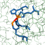
| - | CPA from ''B. taurus'' has | + | As previously stated, CPA from ''B. taurus'' has the ability to bind two Zn<sup>2+</sup> ions in its active site. The binding of only one Zn<sup>2+</sup> ion is [http://en.wikipedia.org/wiki/Catalysis catalytic], while the binding of a second is [http://en.wikipedia.org/wiki/Reaction_inhibitor inhibitory]. These Zn<sup>2+</sup> ions are connected to each other via a hydroxy-bridge (Figure 4) with a distance of 3.48 [http://en.wikipedia.org/wiki/%C3%85ngstr%C3%B6m Å].<ref name="CPA1" /> [[Image:1CPXhydroxybridge.png|150 px|right|thumb|Figure 4: Hydroxy-bridge between catalytic and inhibitory zinc ions. The catalytic Zn<sup>2+</sup> ion (shown in orange on the right) is bridged to the inhibitory Zn<sup>2+</sup> ion (shown in orange on the left) by a OH<sup>-</sup> (shown in red).]] In <scene name='69/694222/3cpas1subsiteglu270/1'>the CPA structure containing only the catalytic Zn<sup>2+</sup> ion</scene> ([http://www.rcsb.org/pdb/explore/explore.do?structureId=3cpa 3CPA]), a water molecule complexed to the zinc is able to be deprotonated by Glu270 to allow for normal initiation of hydrolysis. However, when <scene name='69/694222/Glu270wiz/7'>the inhibitory Zn<sup>2+</sup> ion</scene> is also present ([http://www.rcsb.org/pdb/explore/explore.do?structureId=1cpx 1CPX]), it occupies the physical space that would normally be occupied by the water molecule. Thus, the inhibitory Zn<sup>2+</sup> ion interacts with the carboxylate group of Glu270. The Glu270 now simply stabilizes the second Zn<sup>2+</sup> and is unable to perform its usual base catalyst role while the catalytic Zn<sup>2+</sup> ion (shown in green) is still being stabilized in place by His69, Glu72, and His196 (shown in orange). |
==Other Inhibitors== | ==Other Inhibitors== | ||
Revision as of 23:11, 2 April 2017
| This Sandbox is Reserved from 02/09/2015, through 05/31/2016 for use in the course "CH462: Biochemistry 2" taught by Geoffrey C. Hoops at the Butler University. This reservation includes Sandbox Reserved 1051 through Sandbox Reserved 1080. |
To get started:
More help: Help:Editing |
Carboxypeptidase A in Bos taurus
| |||||||||||
References
- ↑ 1.00 1.01 1.02 1.03 1.04 1.05 1.06 1.07 1.08 1.09 1.10 1.11 1.12 1.13 Bukrinsky JT, Bjerrum MJ, Kadziola A. 1998. Native carboxypeptidase A in a new crystal environment reveals a different conformation of the important tyrosine 248. Biochemistry. 37(47):16555-16564. DOI: 10.1021/bi981678i
- ↑ 2.0 2.1 2.2 2.3 2.4 2.5 2.6 Christianson DW, Lipscomb WN. 1989. Carboxypeptidase A. Acc. Chem. Res. 22:62-69.
- ↑ Suh J, Cho W, Chung S. 1985. Carboxypeptidase A-catalyzed hydrolysis of α-(acylamino)cinnamoyl derivatives of L-β-phenyllactate and L-phenylalaninate: evidence for acyl-enzyme intermediates. J. Am. Chem. Soc. 107:4530-4535. DOI: 10.1021/ja00301a025
- ↑ Geoghegan, KF, Galdes, A, Martinelli, RA, Holmquist, B, Auld, DS, Vallee, BL. 1983. Cryospectroscopy of intermediates in the mechanism of carboxypeptidase A. Biochem. 22(9):2255-2262. DOI: 10.1021/bi00278a031
- ↑ Kaplan, AP, Bartlett, PA. 1991. Synthesis and evaluation of an inhibitor of carboxypeptidase A with a Ki value in the femtomolar range. Biochem. 30(33):8165-8170. PMID: 1868091
- ↑ Worthington, K., Worthington, V. 1993. Worthington Enzyme Manual: Enzymes and Related Biochemicals. Freehold (NJ): Worthington Biochemical Corporation; [2011; accessed March 28, 2017]. Carboxypeptidase A. http://www.worthington-biochem.com/COA/
- ↑ Pitout, MJ, Nel, W. 1969. The inhibitory effect of ochratoxin a on bovine carboxypeptidase a in vitro. Biochem. Pharma. 18(8):1837-1843. DOI: 0.1016/0006-2952(69)90279-2
- ↑ Normant, E, Martres, MP, Schwartz, JC, Gros, C. 1995. Purification, cDNA cloning, functional expression, and characterization of a 26-kDa endogenous mammalian carboxypeptidase inhibitor. Proc. Natl. Acad. Sci. 92(26):12225-12229. PMCID: PMC40329
Student Contributors
- Thomas Baldwin
- Michael Melbardis
- Clay Schnell
Proteopedia Page Contributors and Editors (what is this?)
Michael Melbardis, Douglas Schnell, Thomas Baldwin, Geoffrey C. Hoops, Michal Harel


