This old version of Proteopedia is provided for student assignments while the new version is undergoing repairs. Content and edits done in this old version of Proteopedia after March 1, 2026 will eventually be lost when it is retired in about June of 2026.
Apply for new accounts at the new Proteopedia. Your logins will work in both the old and new versions.
Sandbox Reserved 1240
From Proteopedia
(Difference between revisions)
| Line 2: | Line 2: | ||
==<b>Structure</b>== | ==<b>Structure</b>== | ||
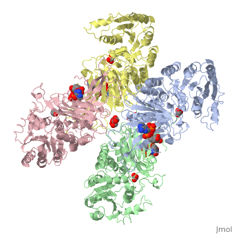
<StructureSection load='1qki' size='340' side='right' caption='Glucose-6-Phosphate Dehydrogenase at a resolution of 3.0 Å' scene=''> | <StructureSection load='1qki' size='340' side='right' caption='Glucose-6-Phosphate Dehydrogenase at a resolution of 3.0 Å' scene=''> | ||
| - | Overall, the structure of Glucose-6-Phosphate Dehydrogenase (G6PD) is a tetramer, composed of a dimer of dimers. Glucose-6-Phosphate Dehydrogenase also features a structural nicotinamide adenine, which is only present in eukaryotes and is rare biologically. Glucose-6-Phosphate Dehydrogenase is 515 amino acids long. | + | Overall, the structure of Glucose-6-Phosphate Dehydrogenase (<i>G6PD</i>) is a tetramer, composed of a dimer of dimers. Glucose-6-Phosphate Dehydrogenase also features a structural nicotinamide adenine, which is only present in eukaryotes and is rare biologically. Glucose-6-Phosphate Dehydrogenase is 515 amino acids long. |
== <b>Function</b> == | == <b>Function</b> == | ||
| Line 15: | Line 15: | ||
Glucose-6-Phosphate Dehydrogenase plays a key role in maintaining homeostasis. Not only does it catalyze the rate limiting step of the oxidative pentose phosphate pathway, it maintains the shape of red blood cells by protecting against potential deadly reactive oxygen species. Glucose-6-Phosphate dehydrogenase deficiency is the most common enzyme deficiency in humans, affecting over 400 million people worldwide. G6PD deficiency resulted in 4100 deaths worldwide in 2013 | Glucose-6-Phosphate Dehydrogenase plays a key role in maintaining homeostasis. Not only does it catalyze the rate limiting step of the oxidative pentose phosphate pathway, it maintains the shape of red blood cells by protecting against potential deadly reactive oxygen species. Glucose-6-Phosphate dehydrogenase deficiency is the most common enzyme deficiency in humans, affecting over 400 million people worldwide. G6PD deficiency resulted in 4100 deaths worldwide in 2013 | ||
| - | + | == <b>Structural highlights </b> == | |
| - | == Structural highlights == | + | The two dimers are held together by charge-charge interactions |
| - | + | ||
| - | + | ||
</StructureSection> | </StructureSection> | ||
== References == | == References == | ||
<references/> | <references/> | ||
Revision as of 14:49, 20 April 2017
| This Sandbox is Reserved from Jan 17 through June 31, 2017 for use in the course Biochemistry II taught by Jason Telford at the Maryville University, St. Louis, USA. This reservation includes Sandbox Reserved 1225 through Sandbox Reserved 1244. |
To get started:
More help: Help:Editing |
Structure
| |||||||||||