User:Rafael Romero Becerra/Sandbox 1
From Proteopedia
(Difference between revisions)
| Line 10: | Line 10: | ||
PCSK9 is the ninth known member of the mammalian subtilisin (S8) serine proprotein convertase (PC) family that carries out the proteolytic maturation of secretory proteins such as neuropeptides, prohormones and cytokines. Humans have nine different PCs that can be divided between S8A and S8B subfamilies. PCSK9 is classified in subfamily S8A <ref name=Piper>DOI 10.1016/j.str.2007.04.004 </ref>. | PCSK9 is the ninth known member of the mammalian subtilisin (S8) serine proprotein convertase (PC) family that carries out the proteolytic maturation of secretory proteins such as neuropeptides, prohormones and cytokines. Humans have nine different PCs that can be divided between S8A and S8B subfamilies. PCSK9 is classified in subfamily S8A <ref name=Piper>DOI 10.1016/j.str.2007.04.004 </ref>. | ||
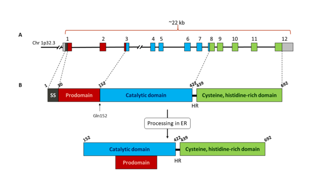
| - | The human gene for PCSK9 is 22 kb length and it is located in chromosome 1p32.3. It contains 11 introns and 12 exons that encode the 692 amino acids of the enzyme. The sequence of the protein is characterized by a signal sequence (amino acids 1-30), a prodomain (amino acids 31-152), and a catalytic domain, followed by a C-terminal region of 243 amino acids which is rich in cysteine and histidine residues | + | The human gene for PCSK9 is 22 kb length and it is located in chromosome 1p32.3. It contains 11 introns and 12 exons that encode the 692 amino acids of the enzyme. The sequence of the protein is characterized by a signal sequence (amino acids 1-30), a prodomain (amino acids 31-152), and a catalytic domain, followed by a C-terminal region of 243 amino acids which is rich in cysteine and histidine residues. PCSK9 is mainly expressed in the liver, intestine and kidney, and it can also be in the nervous system. It is synthesized as a precursor of ~74 kDa that is processed in the endoplasmic reticulum (ER) where it undergoes cleavage of its signal peptide and intramolecular autocatalytic cleavage producing a ~60-kDa catalytic fragment. The autocatalysis of the zymogen takes place between Gln152 and Ser153 <ref name=Naureckiene>PMID:14622975</ref>. This cleavage is necessary for transport from ER to the Golgi body and for secretion. The cleaved prodomain of ~14 kDa remains associated with the catalytic domain, which is unique to PCSK9. This facilitates protein folding, permits the mature protein to move from ER into the secretory pathway and regulates the catalytic activity of the enzyme by blocking the access to the catalytic site <ref name=Abifadel /><ref name=Hess />.[[Image:PCSK9_domains2.png|thumb|center|640x360px| Schematic representation of PCSK9 gene (A) and protein (B). A: exons of PCSK9 gene are shown as coloured boxes. Each colour corresponds to a different domain of the protein. B: Representation of PCSK9 protein domains: signal sequence (SS, black), prodomain (red), catalytic domain (blue) and Cys, His rich C-terminal domain (green). The autocatalytic cleavage site at Gln152 is indicated with an arrow. Once the protein has been processed in the endoplasmic reticulum (ER), the prodomain remains bound to the catalytic domain. The catalytic domain is linked to the C-terminal domain through a 18 amino acids hinge region (HR). Numbers above each domain indicate the amino acid number of the protein sequence.]] |
PCSK9 can be found in plasma in two forms: the mature and secreted form of ~60 kDa, and as an inactivated fragment of ~53 kDa produced by the cleavage of the mature form at the motive RFHR218↓ by other proprotein convertases, mainly furin and/or PC5/6A <ref name=Benjannet>DOI 10.1074/jbc.M606495200</ref>. | PCSK9 can be found in plasma in two forms: the mature and secreted form of ~60 kDa, and as an inactivated fragment of ~53 kDa produced by the cleavage of the mature form at the motive RFHR218↓ by other proprotein convertases, mainly furin and/or PC5/6A <ref name=Benjannet>DOI 10.1074/jbc.M606495200</ref>. | ||
| Line 23: | Line 23: | ||
Although the first role suggested for PCSK9 was neuronal differentiation <ref name=Seidah />, later it was found that PCSK9 is involved in LDL cholesterol metabolism. | Although the first role suggested for PCSK9 was neuronal differentiation <ref name=Seidah />, later it was found that PCSK9 is involved in LDL cholesterol metabolism. | ||
| - | The best-characterized role of the mature and secreted form of PCSK9 (the ~60 kDa cleaved enzyme with the ~14-kDa prodomain associated to the catalytic domain) is targeting LDLRs for degradation in the liver. The catalytic subunit binds the epidermal growth factor-A (EGF-A) domain of the LDLR at the hepatocyte cell surface leading to LDLR internalization and degradation | + | The best-characterized role of the mature and secreted form of PCSK9 (the ~60 kDa cleaved enzyme with the ~14-kDa prodomain associated to the catalytic domain) is targeting LDLRs for degradation in the liver. The catalytic subunit binds the epidermal growth factor-A (EGF-A) domain of the LDLR at the hepatocyte cell surface leading to LDLR internalization and degradation. |
Once LDL cholesterol binds LDLR, it enters the cell through clathrin-coated vesicles. After internalization, the acidic pH of endosomes disrupts the association of LDL cholesterol from its receptor. LDL particles remain within the endosome while a recycling vesicle returns the LDLR to the cell surface. Endosomes containing LDL cholesterol fuse with lysomes where LDL is degraded and cholesterol esters are hydrolyzed. The free cholesterol is then distributed to other cellular compartments. At the hepatocyte cell surface, the catalytic domain of PCSK9 can also bind LDLR. The complex is the internalized via clathrin-coated vesicles. Within the endosome, the affinity of PCSK9 for the LDLR is enhanced due to the low pH, preventing the recycling of the receptor to the cell surface. The complex is then directed to the lysosome, where both components, LDLR and PCSK9, are degraded <ref name=Burke>DOI 10.1146/annurev-pharmtox-010716-104944</ref><ref name=Hess />. In addition, in vitro studies in hepatocytes suggest that PCSK9 might also enhance intracellular LDLR degradation prior to its secretion. When PCSK9 binds to LDLR within the Golgi complex, there is an increase in the traffic of LDLR bound to PCSK9 from the trans Golgi network to lysosomes for degradation, instead of directing the receptors to the cell surface <ref name=Poirier>DOI 10.1074/jbc.M109.037085</ref>. It has been suggested that PCSK9 might also induced LDLR degradation by ubiquitination of the receptor <ref name=Chen>DOI 10.1016/j.bbrc.2011.10.110</ref>. | Once LDL cholesterol binds LDLR, it enters the cell through clathrin-coated vesicles. After internalization, the acidic pH of endosomes disrupts the association of LDL cholesterol from its receptor. LDL particles remain within the endosome while a recycling vesicle returns the LDLR to the cell surface. Endosomes containing LDL cholesterol fuse with lysomes where LDL is degraded and cholesterol esters are hydrolyzed. The free cholesterol is then distributed to other cellular compartments. At the hepatocyte cell surface, the catalytic domain of PCSK9 can also bind LDLR. The complex is the internalized via clathrin-coated vesicles. Within the endosome, the affinity of PCSK9 for the LDLR is enhanced due to the low pH, preventing the recycling of the receptor to the cell surface. The complex is then directed to the lysosome, where both components, LDLR and PCSK9, are degraded <ref name=Burke>DOI 10.1146/annurev-pharmtox-010716-104944</ref><ref name=Hess />. In addition, in vitro studies in hepatocytes suggest that PCSK9 might also enhance intracellular LDLR degradation prior to its secretion. When PCSK9 binds to LDLR within the Golgi complex, there is an increase in the traffic of LDLR bound to PCSK9 from the trans Golgi network to lysosomes for degradation, instead of directing the receptors to the cell surface <ref name=Poirier>DOI 10.1074/jbc.M109.037085</ref>. It has been suggested that PCSK9 might also induced LDLR degradation by ubiquitination of the receptor <ref name=Chen>DOI 10.1016/j.bbrc.2011.10.110</ref>. | ||
Revision as of 17:39, 30 December 2017
| |||||||||||
References
- ↑ 1.0 1.1 Seidah NG, Benjannet S, Wickham L, Marcinkiewicz J, Jasmin SB, Stifani S, Basak A, Prat A, Chretien M. The secretory proprotein convertase neural apoptosis-regulated convertase 1 (NARC-1): liver regeneration and neuronal differentiation. Proc Natl Acad Sci U S A. 2003 Feb 4;100(3):928-33. Epub 2003 Jan 27. PMID:12552133 doi:http://dx.doi.org/10.1073/pnas.0335507100
- ↑ 2.0 2.1 2.2 Abifadel M, Rabes JP, Devillers M, Munnich A, Erlich D, Junien C, Varret M, Boileau C. Mutations and polymorphisms in the proprotein convertase subtilisin kexin 9 (PCSK9) gene in cholesterol metabolism and disease. Hum Mutat. 2009 Apr;30(4):520-9. doi: 10.1002/humu.20882. PMID:19191301 doi:http://dx.doi.org/10.1002/humu.20882
- ↑ 3.0 3.1 3.2 3.3 3.4 3.5 Hess CN, Low Wang CC, Hiatt WR. PCSK9 Inhibitors: Mechanisms of Action, Metabolic Effects, and Clinical Outcomes. Annu Rev Med. 2017 Nov 2. doi: 10.1146/annurev-med-042716-091351. PMID:29095667 doi:http://dx.doi.org/10.1146/annurev-med-042716-091351
- ↑ Piper DE, Jackson S, Liu Q, Romanow WG, Shetterly S, Thibault ST, Shan B, Walker NP. The crystal structure of PCSK9: a regulator of plasma LDL-cholesterol. Structure. 2007 May;15(5):545-52. PMID:17502100 doi:http://dx.doi.org/10.1016/j.str.2007.04.004
- ↑ Naureckiene S, Ma L, Sreekumar K, Purandare U, Lo CF, Huang Y, Chiang LW, Grenier JM, Ozenberger BA, Jacobsen JS, Kennedy JD, DiStefano PS, Wood A, Bingham B. Functional characterization of Narc 1, a novel proteinase related to proteinase K. Arch Biochem Biophys. 2003 Dec 1;420(1):55-67. PMID:14622975
- ↑ Benjannet S, Rhainds D, Hamelin J, Nassoury N, Seidah NG. The proprotein convertase (PC) PCSK9 is inactivated by furin and/or PC5/6A: functional consequences of natural mutations and post-translational modifications. J Biol Chem. 2006 Oct 13;281(41):30561-72. Epub 2006 Aug 15. PMID:16912035 doi:http://dx.doi.org/10.1074/jbc.M606495200
- ↑ Dewpura T, Raymond A, Hamelin J, Seidah NG, Mbikay M, Chretien M, Mayne J. PCSK9 is phosphorylated by a Golgi casein kinase-like kinase ex vivo and circulates as a phosphoprotein in humans. FEBS J. 2008 Jul;275(13):3480-93. doi: 10.1111/j.1742-4658.2008.06495.x. Epub, 2008 May 22. PMID:18498363 doi:http://dx.doi.org/10.1111/j.1742-4658.2008.06495.x
- ↑ 8.0 8.1 Costet P, Cariou B, Lambert G, Lalanne F, Lardeux B, Jarnoux AL, Grefhorst A, Staels B, Krempf M. Hepatic PCSK9 expression is regulated by nutritional status via insulin and sterol regulatory element-binding protein 1c. J Biol Chem. 2006 Mar 10;281(10):6211-8. doi: 10.1074/jbc.M508582200. Epub 2006, Jan 6. PMID:16407292 doi:http://dx.doi.org/10.1074/jbc.M508582200
- ↑ Dubuc G, Chamberland A, Wassef H, Davignon J, Seidah NG, Bernier L, Prat A. Statins upregulate PCSK9, the gene encoding the proprotein convertase neural apoptosis-regulated convertase-1 implicated in familial hypercholesterolemia. Arterioscler Thromb Vasc Biol. 2004 Aug;24(8):1454-9. doi:, 10.1161/01.ATV.0000134621.14315.43. Epub 2004 Jun 3. PMID:15178557 doi:http://dx.doi.org/10.1161/01.ATV.0000134621.14315.43
- ↑ Burke AC, Dron JS, Hegele RA, Huff MW. PCSK9: Regulation and Target for Drug Development for Dyslipidemia. Annu Rev Pharmacol Toxicol. 2017 Jan 6;57:223-244. doi:, 10.1146/annurev-pharmtox-010716-104944. Epub 2016 Aug 8. PMID:27575716 doi:http://dx.doi.org/10.1146/annurev-pharmtox-010716-104944
- ↑ Poirier S, Mayer G, Poupon V, McPherson PS, Desjardins R, Ly K, Asselin MC, Day R, Duclos FJ, Witmer M, Parker R, Prat A, Seidah NG. Dissection of the endogenous cellular pathways of PCSK9-induced low density lipoprotein receptor degradation: evidence for an intracellular route. J Biol Chem. 2009 Oct 16;284(42):28856-64. doi: 10.1074/jbc.M109.037085. Epub, 2009 Jul 27. PMID:19635789 doi:http://dx.doi.org/10.1074/jbc.M109.037085
- ↑ Chen Y, Wang H, Yu L, Yu X, Qian YW, Cao G, Wang J. Role of ubiquitination in PCSK9-mediated low-density lipoprotein receptor degradation. Biochem Biophys Res Commun. 2011 Nov 25;415(3):515-8. doi:, 10.1016/j.bbrc.2011.10.110. Epub 2011 Nov 2. PMID:22074827 doi:10.1016/j.bbrc.2011.10.110
- ↑ Sharotri V, Collier DM, Olson DR, Zhou R, Snyder PM. Regulation of epithelial sodium channel trafficking by proprotein convertase subtilisin/kexin type 9 (PCSK9). J Biol Chem. 2012 Jun 1;287(23):19266-74. doi: 10.1074/jbc.M112.363382. Epub 2012, Apr 9. PMID:22493497 doi:10.1074/jbc.M112.363382
- ↑ Hanson, R. M., Prilusky, J., Renjian, Z., Nakane, T. and Sussman, J. L. (2013), JSmol and the Next-Generation Web-Based Representation of 3D Molecular Structure as Applied to Proteopedia. Isr. J. Chem., 53:207-216. doi:http://dx.doi.org/10.1002/ijch.201300024
- ↑ Herraez A. Biomolecules in the computer: Jmol to the rescue. Biochem Mol Biol Educ. 2006 Jul;34(4):255-61. doi: 10.1002/bmb.2006.494034042644. PMID:21638687 doi:10.1002/bmb.2006.494034042644
- ↑ 16.0 16.1 16.2 16.3 16.4 16.5 El Khoury P, Elbitar S, Ghaleb Y, Khalil YA, Varret M, Boileau C, Abifadel M. PCSK9 Mutations in Familial Hypercholesterolemia: from a Groundbreaking Discovery to Anti-PCSK9 Therapies. Curr Atheroscler Rep. 2017 Oct 17;19(12):49. doi: 10.1007/s11883-017-0684-8. PMID:29038906 doi:http://dx.doi.org/10.1007/s11883-017-0684-8
- ↑ 17.0 17.1 Zhang Y, Ultsch M, Skelton NJ, Burdick DJ, Beresini MH, Li W, Kong-Beltran M, Peterson A, Quinn J, Chiu C, Wu Y, Shia S, Moran P, Di Lello P, Eigenbrot C, Kirchhofer D. Discovery of a cryptic peptide-binding site on PCSK9 and design of antagonists. Nat Struct Mol Biol. 2017 Aug 21. doi: 10.1038/nsmb.3453. PMID:28825733 doi:http://dx.doi.org/10.1038/nsmb.3453
- ↑ Giunzioni I, Tavori H. New developments in atherosclerosis: clinical potential of PCSK9 inhibition. Vasc Health Risk Manag. 2015 Aug 24;11:493-501. doi: 10.2147/VHRM.S74692., eCollection 2015. PMID:26345307 doi:http://dx.doi.org/10.2147/VHRM.S74692
- ↑ Mitchell T, Chao G, Sitkoff D, Lo F, Monshizadegan H, Meyers D, Low S, Russo K, DiBella R, Denhez F, Gao M, Myers J, Duke G, Witmer M, Miao B, Ho SP, Khan J, Parker RA. Pharmacologic Profile of the Adnectin BMS-962476, a Small Protein Biologic Alternative to PCSK9 Antibodies for LDL Lowering. J Pharmacol Exp Ther. 2014 Jun 10. pii: jpet.114.214221. PMID:24917546 doi:http://dx.doi.org/10.1124/jpet.114.214221
- ↑ Mullard A. Nine paths to PCSK9 inhibition. Nat Rev Drug Discov. 2017 Apr 28;16(5):299-301. doi: 10.1038/nrd.2017.83. PMID:28450722 doi:http://dx.doi.org/10.1038/nrd.2017.83

