This old version of Proteopedia is provided for student assignments while the new version is undergoing repairs. Content and edits done in this old version of Proteopedia after March 1, 2026 will eventually be lost when it is retired in about June of 2026.
Apply for new accounts at the new Proteopedia. Your logins will work in both the old and new versions.
Sandbox Reserved 1329
From Proteopedia
(Difference between revisions)
| Line 10: | Line 10: | ||
== Relevance == | == Relevance == | ||
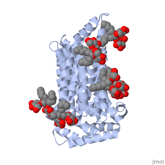
| - | Both the amino and carbonyl termini of the protein are exposed on the cytoplasmic side of the plasma membrane. This protein functions via the | + | Both the amino and carbonyl termini of the protein are exposed on the cytoplasmic side of the plasma membrane. This protein functions via the alternative confirmation model. A transport protein exposes a substrate towards either the outside or inside the cell. When the glucose or hexose binds to the site, it catalyzes a conformational change, releasing the glucose on the other side of the membrane. GLUT3 is a unique glucose transporter in that it functions even in times of low glucose. |
== Structural highlights == | == Structural highlights == | ||
| - | The protein contains 12 membrane-spanning alpha helices and has no known post-translational modifications. The first 6 transmembrane helices are in a pseudo symmetrical configuration relative to the last 6 helices. Helices 1, 2, 4, 5, 7, 8, 10, and 11 form an inner bundle that is stabilized by the outer helices 3, 6, 9, and 12. The GLUT3 protein is comprised of ~500 amino acid residues. It has a single site for N-Linked glycosylation, a central cytoplasmic linker domain, and exhibit topologies with their N and C termini, which are both positioned in the cytoplasm. | + | The protein contains <scene name='77/777649/Helices/1'>12 membrane-spanning alpha helices</scene> and has no known post-translational modifications. The first 6 transmembrane helices are in a pseudo symmetrical configuration relative to the last 6 helices. Helices 1, 2, 4, 5, 7, 8, 10, and 11 form an inner bundle that is stabilized by the outer helices 3, 6, 9, and 12. The GLUT3 protein is comprised of ~500 amino acid residues. It has a single site for N-Linked glycosylation, a central cytoplasmic linker domain, and exhibit topologies with their N and C termini, which are both positioned in the cytoplasm. |
The protein also contains nine <scene name='77/777649/Ligands_37x/1'>37X ligands</scene>. The molecule itself is octyl glucose neopentyl glycol. | The protein also contains nine <scene name='77/777649/Ligands_37x/1'>37X ligands</scene>. The molecule itself is octyl glucose neopentyl glycol. | ||
Revision as of 19:02, 27 February 2018
| This Sandbox is Reserved from January through July 31, 2018 for use in the course HLSC322: Principles of Genetics and Genomics taught by Genevieve Houston-Ludlam at the University of Maryland, College Park, USA. This reservation includes Sandbox Reserved 1311 through Sandbox Reserved 1430. |
To get started:
More help: Help:Editing |
Human Glucose Transporter GLUT3/SLC2A3
| |||||||||||
References
https://www.ncbi.nlm.nih.gov/pubmed/19690067
https://www.ncbi.nlm.nih.gov/books/NBK6545/
https://www.ncbi.nlm.nih.gov/pmc/articles/PMC4104978/
Carruthers A, DeZutter J, Ganguly A, Devaskar SU. Will the original glucose transporter isoform please stand up! Am J Physiol Endocrinol Metab. 2009;297(4):E836-848.
