This old version of Proteopedia is provided for student assignments while the new version is undergoing repairs. Content and edits done in this old version of Proteopedia after March 1, 2026 will eventually be lost when it is retired in about June of 2026.
Apply for new accounts at the new Proteopedia. Your logins will work in both the old and new versions.
Biotin Protein Ligase
From Proteopedia
(Difference between revisions)
| Line 7: | Line 7: | ||
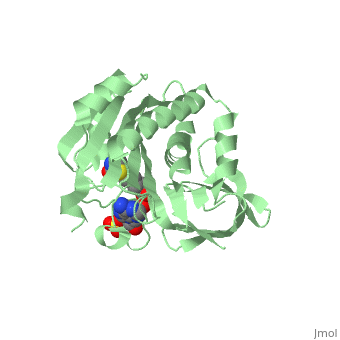
Biotinylation is catalysed through a two-step reaction where biotin is first activated to biotinyl-5′-AMP in an ATP dependent manner. The biotin is then transferred onto the ε-amino group of a specific target lysine residue. The reaction mechanism is related to that of amino acyl-tRNA synthetases and lipoyl ligases where the reaction proceeds through the formation of an adenylated intermediate, suggesting a common ancestral relationship <ref>pmid 18442489</ref> . | Biotinylation is catalysed through a two-step reaction where biotin is first activated to biotinyl-5′-AMP in an ATP dependent manner. The biotin is then transferred onto the ε-amino group of a specific target lysine residue. The reaction mechanism is related to that of amino acyl-tRNA synthetases and lipoyl ligases where the reaction proceeds through the formation of an adenylated intermediate, suggesting a common ancestral relationship <ref>pmid 18442489</ref> . | ||
| - | Of all the BPL’s, E.coli (BirA) is by far the most characterised and understood family member. A recent ensemble of BPL structures from the thermophilic archea Pirococcus Horikoshii OT3 <ref>pmid 16510991</ref> have also provided new insights into the catalytic mechanism of BPLs. | + | Of all the BPL’s, E.coli ('''BirA''') is by far the most characterised and understood family member. A recent ensemble of BPL structures from the thermophilic archea Pirococcus Horikoshii OT3 <ref>pmid 16510991</ref> have also provided new insights into the catalytic mechanism of BPLs. |
== Structural highlights == | == Structural highlights == | ||
Revision as of 11:50, 4 March 2018
| |||||||||||
3D structures of Biotin Protein Ligase
Updated on 04-March-2018
References
- ↑ Pendini NR, Bailey LM, Booker GW, Wilce MC, Wallace JC, Polyak SW. Microbial biotin protein ligases aid in understanding holocarboxylase synthetase deficiency. Biochim Biophys Acta. 2008 Jul-Aug;1784(7-8):973-82. Epub 2008 Apr 9. PMID:18442489 doi:10.1016/j.bbapap.2008.03.011
- ↑ Bagautdinov B, Kuroishi C, Sugahara M, Kunishima N. Purification, crystallization and preliminary crystallographic analysis of the biotin-protein ligase from Pyrococcus horikoshii OT3. Acta Crystallogr Sect F Struct Biol Cryst Commun. 2005 Feb 1;61(Pt, 2):193-5. Epub 2005 Jan 8. PMID:16510991 doi:10.1107/S1744309104034360
- ↑ Wilson KP, Shewchuk LM, Brennan RG, Otsuka AJ, Matthews BW. Escherichia coli biotin holoenzyme synthetase/bio repressor crystal structure delineates the biotin- and DNA-binding domains. Proc Natl Acad Sci U S A. 1992 Oct 1;89(19):9257-61. PMID:1409631
Proteopedia Page Contributors and Editors (what is this?)
Michal Harel, Nicole R Pendini, Alexander Berchansky, David Canner, Jaime Prilusky
