This old version of Proteopedia is provided for student assignments while the new version is undergoing repairs. Content and edits done in this old version of Proteopedia after March 1, 2026 will eventually be lost when it is retired in about June of 2026.
Apply for new accounts at the new Proteopedia. Your logins will work in both the old and new versions.
Arginine kinase
From Proteopedia
(Difference between revisions)
| Line 13: | Line 13: | ||
== Structural highlights == | == Structural highlights == | ||
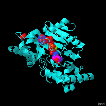
| - | The <scene name='71/715906/Cv/3'>active site</scene> of AK is located between its N-terminal helical region and the larger C-terminal region.<ref>PMID:9671698</ref> | + | The <scene name='71/715906/Cv/3'>active site</scene> of AK is located between its N-terminal helical region and the larger C-terminal region. <ref>PMID:9671698</ref> Water molecules are shown as red spheres. |
</StructureSection> | </StructureSection> | ||
Revision as of 10:52, 8 May 2018
| |||||||||||
3D structures of arginine kinase
Updated on 08-May-2018
References
- ↑ Wang Z, Qiao Z, Ye S, Zhang R. Structure of a double-domain phosphagen kinase reveals an asymmetric arrangement of the tandem domains. Acta Crystallogr D Biol Crystallogr. 2015 Apr;71(Pt 4):779-89. doi:, 10.1107/S1399004715001169. Epub 2015 Mar 26. PMID:25849389 doi:http://dx.doi.org/10.1107/S1399004715001169
- ↑ Zhou G, Somasundaram T, Blanc E, Parthasarathy G, Ellington WR, Chapman MS. Transition state structure of arginine kinase: implications for catalysis of bimolecular reactions. Proc Natl Acad Sci U S A. 1998 Jul 21;95(15):8449-54. PMID:9671698
