User:Mark Hoelzer/Sandbox1
From Proteopedia
(Difference between revisions)
| Line 6: | Line 6: | ||
| - | { | + | {| class="wikitable" |
| - | + | |- | |
| - | + | ! Header 1 | |
| - | + | ! Header 2 | |
| + | ! Header 3 | ||
| + | |- | ||
| + | | row 1, cell 1 | ||
| + | | row 1, cell 2 | ||
| + | | row 1, cell 3 | ||
| + | |- | ||
| + | | row 2, cell 1 | ||
| + | | row 2, cell 2 | ||
| + | | row 2, cell 3 | ||
| + | |} | ||
| + | |||
| + | |||
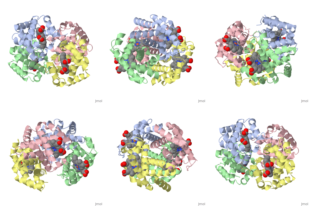
<StructureSection load='1a3n' side='left' size='400' caption='Hemoglobin based on 1a3n.pdb' scene=''> | <StructureSection load='1a3n' side='left' size='400' caption='Hemoglobin based on 1a3n.pdb' scene=''> | ||
Revision as of 14:41, 3 April 2018
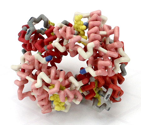
3D Printed Physical Model of Hemoglobin
Shown below is a physical 3D printed model of Hemoglobin, based on the structure 1a3n.pdb. The two alpha-globin chains are colored light red, the two beta globin chains are colored dark red, and the four heme groups are colored yellow.
| Header 1 | Header 2 | Header 3 |
|---|---|---|
| row 1, cell 1 | row 1, cell 2 | row 1, cell 3 |
| row 2, cell 1 | row 2, cell 2 | row 2, cell 3 |
| |||||||||||


