|
Introduction
Threonyl t-RNA Synthetase or Threonyl-tRNA ligase or TARS is a homodimer (150kDa in bacteria and 170kDa in human) and is classified as a class II Aminoacyl-tRNA synthetase enzymes. These ancient enzymes primary function are to add the respective amino acid to the respective transfer Ribonucleic Acid (tRNA-AA), a necessity prep for the protein synthesis pathway [1]. As the name implies, TARS function is to add Threonine amino acid (Thr) to threonine specific tRNA (tRNA-thr) in the presence of Adenosine triphosphate (ATP) and diavalent metal cation. Below displays the overview of TARS Aminoacylation rxn [2].  Overall TARS protein rxn. Substrates includes ATP, Thr and tRNA-thr.
Mechanism
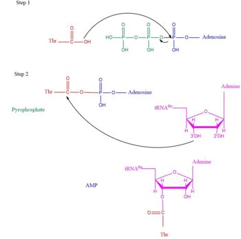
 Arrow pushing of Aminoacylation rxn.[3] TARS adds amino acid to tRNA by a two-step mechanism. First the enzyme binds to both and in the catalytic domain to perform an adenylation reaction in which threonyl adenylate (Thr-AMP) is formed and pyrophosphate (PPi) is released as a byproduct. The image to the right displays the binding of adenylate product to the TARS enzyme (PDB entry 1nyq). This is then follow up by a transferring Thr from Adenosine monophosphate (AMP) molecule to 3'OH site of tRNA-thr. [4] The image to the above demonstrates the arrow pushing occurring to generate threonine bound tRNA-thr.
Disease Relevance
Lately the aaRS family was found to have more function than just aminoacylation. For instance, many aaRs molecules have been found to link with angiogenesis, blood vessel growth occuring within a cancer environment[5]. This is seen in human exogenous TARS in its ability to generate blood vessels within ovarian cancer environment[6]. Studies on the structure of TARS bound to BC194, derivative to the natural antibiotic Borrelidin, were investigated to understand how the angiogenic signaling from TARS occurs[7].
Structural highlights
As mentioned earlier, TARS is homodimer protein found in two forms of a cell, mitochondrial and cytoplasmic. Most of the structures seen will primarly be cytoplasmic. The protein is mainly classified as a alpha beta protein in both bacteria and eukaryotic cells [8]. Each chain is composed of 4 domain classified by SCOP: a N1 domain, a N2 domain, a catalytic domain and an anti-codon domain [9].
N-terminus (Residue 1-241)
The N1-Domain and N-2 domain is the found in the N-terminus of the TARS enzyme chain. Specific N1 is found from 1-62 residue while N2 domain is found from 62-241 residue(PDB: 1TJE). Although located in neighboring regions, the N1-domain and N2-domain deviate dramatically from one another. Structurally, N1-domain has a TGS-fold (short for TARS, GTPase, Spo1 domain as their the most common proteins that present this fold)[10] which has 3 antiparallel beta sheets interacting with an alpha helix causing a formation of roll-like structure (similar to the description by Cath: Ubiquitin-like roll)[11]. The structure has no known function but it is pronounced structure found common within TARS.
This is the opposite of the N2-domain, or sometimes called TARS additional domain. This is ironic as this addition is very important for proofreading TARS activity[12]. TARS N2-domain is described as 2-layer alpha-beta sandwich comprised of mostly alpha helices. Interesting enough, the superfamily of this motif is found to be TARS and Alanyl-tRNA synthetase common domain. This domain function similar by discriminating Threonine and Alanine from Serine amino acid (Ser) as serine is similar structure to both amino acids. The editing domain functions first by moving Ser bound tRNA-Thr from the catalytic domain to the editing site by breaking the bonds nucleotide A73, C74, and C75 from the catalytic site allowing the acceptor arm of tRNA-Thr to flip to the editing domain of S12-S13 site. Research on editing hydrolysis propose that residue Tyr 103 plays an important role of guiding acceptor arm towards the editing domain. The Ser bound to tRNA is hydrolzed by a water molecule, interacting with His73, acting as a nucleophile to the alpha carbon of serine, follow by protonation by a 2nd water molecule interacting with carbonyl of Met181 and amide side chain Lys156. The fidility mechanism of AA-tRNA binding is much similar to alanine tRNA-ligase found in the C-terminus as they share 40% similarity in residues. The subtle difference of the isoforms mitochondrial TARS from cytoplasmic TARS appears in editing as the mitochondrial TARS doesn't have N2-domain and requires interaction to hydrolyze serine [13].
Catalytic Domain (Residue 243-
Anti-Codon Domain
Evolutionary related proteins
List to available structures
References
- ↑ Rajendran V, Kalita P, Shukla H, Kumar A, Tripathi T. Aminoacyl-tRNA synthetases: Structure, function, and drug discovery. Int J Biol Macromol. 2018 May;111:400-414. doi: 10.1016/j.ijbiomac.2017.12.157., Epub 2018 Jan 3. PMID:29305884 doi:http://dx.doi.org/10.1016/j.ijbiomac.2017.12.157
- ↑ Rajendran V, Kalita P, Shukla H, Kumar A, Tripathi T. Aminoacyl-tRNA synthetases: Structure, function, and drug discovery. Int J Biol Macromol. 2018 May;111:400-414. doi: 10.1016/j.ijbiomac.2017.12.157., Epub 2018 Jan 3. PMID:29305884 doi:http://dx.doi.org/10.1016/j.ijbiomac.2017.12.157
- ↑ Lehninger, A. L., Nelson, D. L., & Cox, M. M. (2000). Lehninger principles of biochemistry. New York: Worth Publishers.
- ↑ Lehninger, A. L., Nelson, D. L., & Cox, M. M. (2000). Lehninger principles of biochemistry. New York: Worth Publishers.
- ↑ Mirando AC, Francklyn CS, Lounsbury KM. Regulation of angiogenesis by aminoacyl-tRNA synthetases. Int J Mol Sci. 2014 Dec 19;15(12):23725-48. doi: 10.3390/ijms151223725. PMID:25535072 doi:http://dx.doi.org/10.3390/ijms151223725
- ↑ Williams TF, Mirando AC, Wilkinson B, Francklyn CS, Lounsbury KM. Secreted Threonyl-tRNA synthetase stimulates endothelial cell migration and angiogenesis. Sci Rep. 2013;3:1317. doi: 10.1038/srep01317. PMID:23425968 doi:http://dx.doi.org/10.1038/srep01317
- ↑ Mirando AC, Fang P, Williams TF, Baldor LC, Howe AK, Ebert AM, Wilkinson B, Lounsbury KM, Guo M, Francklyn CS. Aminoacyl-tRNA synthetase dependent angiogenesis revealed by a bioengineered macrolide inhibitor. Sci Rep. 2015 Aug 14;5:13160. doi: 10.1038/srep13160. PMID:26271225 doi:http://dx.doi.org/10.1038/srep13160
- ↑ Teng M, Hilgers MT, Cunningham ML, Borchardt A, Locke JB, Abraham S, Haley G, Kwan BP, Hall C, Hough GW, Shaw KJ, Finn J. Identification of bacteria-selective threonyl-tRNA synthetase substrate inhibitors by structure-based design. J Med Chem. 2013 Feb 28;56(4):1748-60. doi: 10.1021/jm301756m. Epub 2013 Feb 12. PMID:23362938 doi:10.1021/jm301756m
- ↑ Torres-Larios A, Sankaranarayanan R, Rees B, Dock-Bregeon AC, Moras D. Conformational movements and cooperativity upon amino acid, ATP and tRNA binding in threonyl-tRNA synthetase. J Mol Biol. 2003 Aug 1;331(1):201-11. PMID:12875846
- ↑ Wolf YI, Aravind L, Grishin NV, Koonin EV. Evolution of aminoacyl-tRNA synthetases--analysis of unique domain architectures and phylogenetic trees reveals a complex history of horizontal gene transfer events. Genome Res. 1999 Aug;9(8):689-710. PMID:10447505
- ↑ http://www.rcsb.org/pdb/explore/macroMoleculeData.do?structureId=1TJE
- ↑ Dock-Bregeon AC, Rees B, Torres-Larios A, Bey G, Caillet J, Moras D. Achieving error-free translation; the mechanism of proofreading of threonyl-tRNA synthetase at atomic resolution. Mol Cell. 2004 Nov 5;16(3):375-86. PMID:15525511 doi:http://dx.doi.org/10.1016/j.molcel.2004.10.002
- ↑ Ling J, Peterson KM, Simonovic I, Soll D, Simonovic M. The mechanism of pre-transfer editing in yeast mitochondrial threonyl-tRNA synthetase. J Biol Chem. 2012 Aug 17;287(34):28518-25. doi: 10.1074/jbc.M112.372920. Epub, 2012 Jul 6. PMID:22773845 doi:http://dx.doi.org/10.1074/jbc.M112.372920
This is a sample scene created with SAT to by Group, and another to make of the protein. You can make your own scenes on SAT starting from scratch or loading and editing one of these sample scenes.
|


