We apologize for Proteopedia being slow to respond. For the past two years, a new implementation of Proteopedia has been being built. Soon, it will replace this 18-year old system. All existing content will be moved to the new system at a date that will be announced here.
Fibrinogen
From Proteopedia
(Difference between revisions)
| Line 8: | Line 8: | ||
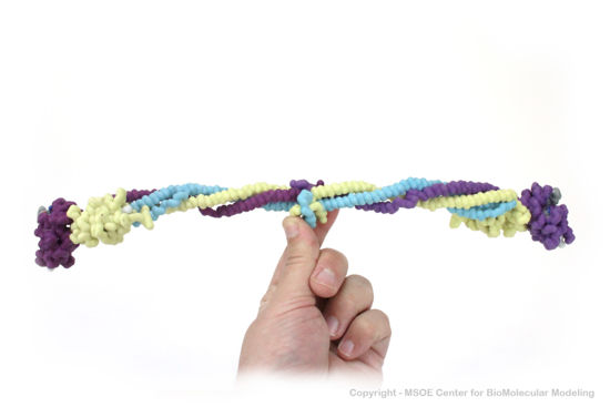
Shown below is a 3D printed physical model of Fibrinogen. The structure is shown as an alpha carbon backbone colored by chain, with the three chains of each copy of fibrinogen colored yellow, blue and purple. | Shown below is a 3D printed physical model of Fibrinogen. The structure is shown as an alpha carbon backbone colored by chain, with the three chains of each copy of fibrinogen colored yellow, blue and purple. | ||
| + | |||
[[Image:fibrinogen1_centerForBioMolecularModeling.jpg|550px]] | [[Image:fibrinogen1_centerForBioMolecularModeling.jpg|550px]] | ||
Revision as of 19:01, 31 July 2019
| |||||||||||
3D Structure of Fibrinogen
Updated on 31-July-2019
References
- ↑ Betts L, Merenbloom BK, Lord ST. The structure of fibrinogen fragment D with the 'A' knob peptide GPRVVE. J Thromb Haemost. 2006 May;4(5):1139-41. PMID:16689770 doi:10.1111/j.1538-7836.2006.01902.x
- ↑ Muszbek L, Bagoly Z, Bereczky Z, Katona E. The involvement of blood coagulation factor XIII in fibrinolysis and thrombosis. Cardiovasc Hematol Agents Med Chem. 2008 Jul;6(3):190-205. PMID:18673233
Proteopedia Page Contributors and Editors (what is this?)
Michal Harel, David Canner, Mark Hoelzer, Alexander Berchansky, Marius Mihasan, Angel Herraez


