We apologize for Proteopedia being slow to respond. For the past two years, a new implementation of Proteopedia has been being built. Soon, it will replace this 18-year old system. All existing content will be moved to the new system at a date that will be announced here.
Nitric Oxide Synthase
From Proteopedia
(Difference between revisions)
| Line 7: | Line 7: | ||
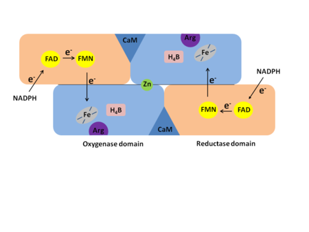
In mammals three isozymes of NOS have been identified: Neuronal NOS (nNOS), inducible NOS (iNOS), and endothelial NOS (eNOS)<ref>Flemming, I., Chapter 3, Biology of Nitric Oxide Synthases, from Microcirculation, Editors Ronald F. Tuma, R. F., Durán, W. N., and Ley, K., 2nd edition, Academic Press, 2008</ref>. (The NOS enzymes are found in numeral organisms. Most facts used here are from the human NOS, but sites from different organisms are used). Neuronal NOS is producing NO in the nervous tissue in both the peripheral and the central nervous system. nNOS is functioning in cell signaling and communication - a vital part of the nervous tissue. Inducible NOS is connected with the immune system. Endothelial NOS is controlling the amount of NO signaling in the endothelial cells eg. blood vessel dilation. An overview of the structural organization of the NOS homodimer is given below. All cofactors are included and the electron transfer pathway which takes place in NOS is indicated. | In mammals three isozymes of NOS have been identified: Neuronal NOS (nNOS), inducible NOS (iNOS), and endothelial NOS (eNOS)<ref>Flemming, I., Chapter 3, Biology of Nitric Oxide Synthases, from Microcirculation, Editors Ronald F. Tuma, R. F., Durán, W. N., and Ley, K., 2nd edition, Academic Press, 2008</ref>. (The NOS enzymes are found in numeral organisms. Most facts used here are from the human NOS, but sites from different organisms are used). Neuronal NOS is producing NO in the nervous tissue in both the peripheral and the central nervous system. nNOS is functioning in cell signaling and communication - a vital part of the nervous tissue. Inducible NOS is connected with the immune system. Endothelial NOS is controlling the amount of NO signaling in the endothelial cells eg. blood vessel dilation. An overview of the structural organization of the NOS homodimer is given below. All cofactors are included and the electron transfer pathway which takes place in NOS is indicated. | ||
{{Clear}} | {{Clear}} | ||
| - | [[Image:schematic_NOS.png | + | [[Image:schematic_NOS.png|left|thumb|450px]] |
{{Clear}} | {{Clear}} | ||
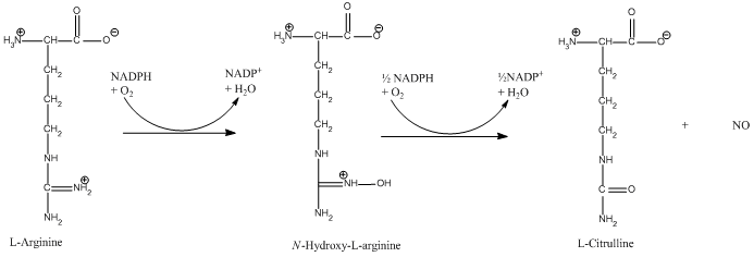
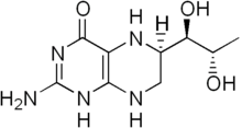
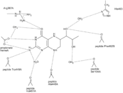
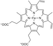
Each NOS monomer is composed of two domains: an N-terminal oxygenase domain and a C-terminal reductase domain. Each subunit is held together by a zinc ion, which is bound by two cysteines from each oxygenase domain. Calmodulin binds to a linker region between the two domains and is necessary for activity. The reductase domain supplies electrons for the NOS reaction which takes place in the oxygenase domain. The reductase domain contains two redox-active prosthetic groups, flavin adenine dinucleotide (FAD) and Flavin mononucleotide (FMN). Nicotinamide adenine dinucleotide phosphate(NADPH) binds to the domain and passes on an electron to FAD which passes the electron on to FMN. FMN passes the electron on to the heme in the oxygenase domain on the opposite subunit. The oxygenase domain contains 5,6,7,8-tetrahydrobiopterin (H<sub>4</sub>B) and the already mentioned heme. These two are also redox active groups. Besides Heme and H<sub>4</sub>B, the oxygenase domain binds the substrate L-arginine which takes part in the NO synthase reaction (see below). | Each NOS monomer is composed of two domains: an N-terminal oxygenase domain and a C-terminal reductase domain. Each subunit is held together by a zinc ion, which is bound by two cysteines from each oxygenase domain. Calmodulin binds to a linker region between the two domains and is necessary for activity. The reductase domain supplies electrons for the NOS reaction which takes place in the oxygenase domain. The reductase domain contains two redox-active prosthetic groups, flavin adenine dinucleotide (FAD) and Flavin mononucleotide (FMN). Nicotinamide adenine dinucleotide phosphate(NADPH) binds to the domain and passes on an electron to FAD which passes the electron on to FMN. FMN passes the electron on to the heme in the oxygenase domain on the opposite subunit. The oxygenase domain contains 5,6,7,8-tetrahydrobiopterin (H<sub>4</sub>B) and the already mentioned heme. These two are also redox active groups. Besides Heme and H<sub>4</sub>B, the oxygenase domain binds the substrate L-arginine which takes part in the NO synthase reaction (see below). | ||
Revision as of 11:44, 8 July 2018
| |||||||||||
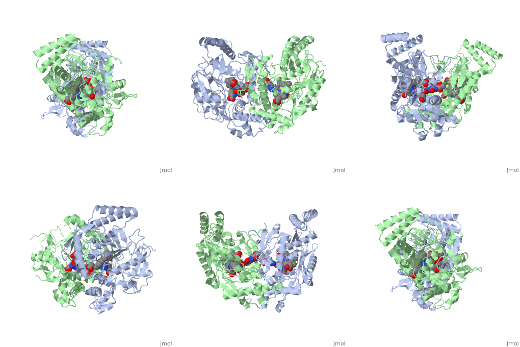
3D Structures of Nitric Oxide Synthase
Updated on 08-July-2018
References
- ↑ Flemming, I., Chapter 3, Biology of Nitric Oxide Synthases, from Microcirculation, Editors Ronald F. Tuma, R. F., Durán, W. N., and Ley, K., 2nd edition, Academic Press, 2008
- ↑ Gorren AC, Mayer B. Nitric-oxide synthase: a cytochrome P450 family foster child. Biochim Biophys Acta. 2007 Mar;1770(3):432-45. Epub 2006 Sep 1. PMID:17014963 doi:10.1016/j.bbagen.2006.08.019
- ↑ Fan B, Wang J, Stuehr DJ, Rousseau DL. NO synthase isozymes have distinct substrate binding sites. Biochemistry. 1997 Oct 21;36(42):12660-5. PMID:9376373 doi:http://dx.doi.org/10.1021/bi9715369
- ↑ Rousseau DL, Li D, Couture M, Yeh SR. Ligand-protein interactions in nitric oxide synthase. J Inorg Biochem. 2005 Jan;99(1):306-23. PMID:15598509 doi:http://dx.doi.org/10.1016/j.jinorgbio.2004.11.007
- ↑ 5.0 5.1 Raman CS, Li H, Martasek P, Kral V, Masters BS, Poulos TL. Crystal structure of constitutive endothelial nitric oxide synthase: a paradigm for pterin function involving a novel metal center. Cell. 1998 Dec 23;95(7):939-50. PMID:9875848
- ↑ 6.0 6.1 6.2 Wei CC, Wang ZQ, Meade AL, McDonald JF, Stuehr DJ. Why do nitric oxide synthases use tetrahydrobiopterin? J Inorg Biochem. 2002 Sep 20;91(4):618-24. PMID:12237227
- ↑ Li H, Igarashi J, Jamal J, Yang W, Poulos TL. Structural studies of constitutive nitric oxide synthases with diatomic ligands bound. J Biol Inorg Chem. 2006 Sep;11(6):753-68. Epub 2006 Jun 28. PMID:16804678 doi:http://dx.doi.org/10.1007/s00775-006-0123-8
- ↑ 8.0 8.1 Fischmann TO, Hruza A, Niu XD, Fossetta JD, Lunn CA, Dolphin E, Prongay AJ, Reichert P, Lundell DJ, Narula SK, Weber PC. Structural characterization of nitric oxide synthase isoforms reveals striking active-site conservation. Nat Struct Biol. 1999 Mar;6(3):233-42. PMID:10074942 doi:http://dx.doi.org/10.1038/6675
- ↑ Crane BR, Rosenfeld RJ, Arvai AS, Ghosh DK, Ghosh S, Tainer JA, Stuehr DJ, Getzoff ED. N-terminal domain swapping and metal ion binding in nitric oxide synthase dimerization. EMBO J. 1999 Nov 15;18(22):6271-81. PMID:10562539 doi:http://dx.doi.org/10.1093/emboj/18.22.6271
- ↑ Garcin ED, Bruns CM, Lloyd SJ, Hosfield DJ, Tiso M, Gachhui R, Stuehr DJ, Tainer JA, Getzoff ED. Structural basis for isozyme-specific regulation of electron transfer in nitric-oxide synthase. J Biol Chem. 2004 Sep 3;279(36):37918-27. Epub 2004 Jun 17. PMID:15208315 doi:http://dx.doi.org/10.1074/jbc.M406204200
Proteopedia Page Contributors and Editors (what is this?)
Sara Toftegaard Petersen, Michal Harel, Mathilde Thomsen, Michael Skovbo Windahl, Alexander Berchansky, Mette Trauelsen, Ann Taylor, Jaime Prilusky, Eran Hodis, Karl Oberholser
