Main Page

From Proteopedia

(Difference between revisions)
Jump to: navigation, search
Line 28: Line 28:
<tr style="font-size: 1.2em; text-align: center;">
-
<td style="padding: 10px;>How to author pages and contribute to Proteopedia</td>
+
<td style="padding: 10px;>[[http://proteopedia.org/w/Help:Contents#For_authors:_contributing_content|How to author pages and contribute to Proteopedia]]</td>
<td style="padding: 10px;></td>
<td style="padding: 10px;>How to get an Interactive 3D Complement for your paper</td>

Revision as of 13:24, 18 October 2018

Because life has more than 2D, Proteopedia helps to understand relationships between structure and function. Proteopedia is a free, collaborative 3D-encyclopedia of proteins & other molecules. ISSN 2310-6301

Selected Pages Art on Science Journals Education
About this image
Coronavirus Spike Protein Priming

by Eric Martz
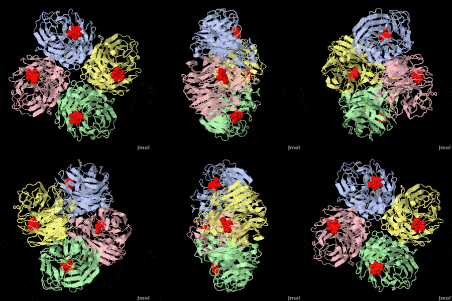
Coronavirus SARS-CoV-2 (responsible for COVID-19) has a spike protein on its surface, which enables it to infect host cells. Initially, proteases in the lungs clip the homo-trimeric spike protein at a unique sequence. This primes it, causing it to extend its receptor binding surface (shown in the above animation), optimizing binding to the host cell's ACE2 receptor (not shown). Next, spike protein initiates fusion of the virus and host cell membranes (not shown), enabling the virus RNA to enter the cell and initiate production of new virions. Knowledge of spike protein's molecular structure and function is crucial to developing effective therapies and vaccines.
>>> Visit this page >>>

About this image
Molecular Sculpture

by Eric Martz
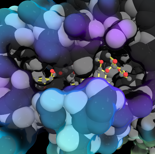
A historical review on sculptures and physical models of macromolecules.

>>> Visit this page >>>

About this image
Interconversion of the specificities of human lysosomal enzymes associated with Fabry and Schindler diseases.

IB Tomasic, MC Metcalf, AI Guce, NE Clark, SC Garman. J. Biol. Chem. 2010 doi: 10.1074/jbc.M110.118588
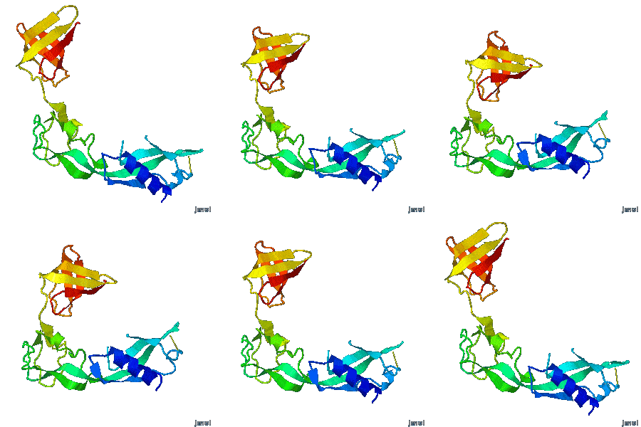
The human lysosomal enzymes α-galactosidase and α-N-acetylgalactosaminidase share 46% amino acid sequence identity and have similar folds. Using a rational protein engineering approach, we interconverted the enzymatic specificity of α-GAL and α-NAGAL. The engineered α-GAL retains the antigenicity but has acquired the enzymatic specificity of α-NAGAL. Conversely, the engineered α-NAGAL retains the antigenicity but has acquired the enzymatic specificity of the α-GAL enzyme. Comparison of the crystal structures of the designed enzyme to the wild-type enzymes shows that active sites superimpose well, indicating success of the rational design. The designed enzymes might be useful as non-immunogenic alternatives in enzyme replacement therapy for treatment of lysosomal storage disorders such as Fabry disease.

>>> Visit this I3DC complement >>>

About this image
Tutorial: The Ramachandran principle, phi (φ) and psi (ψ) angles in proteins

by Eric Martz
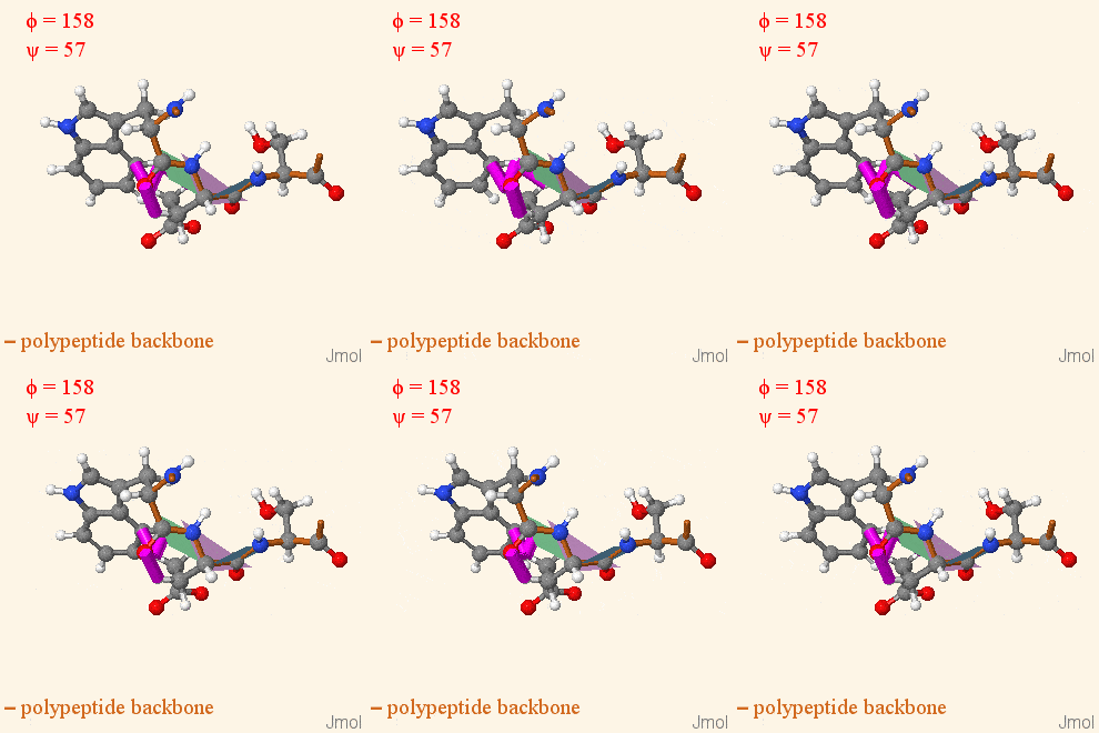
The Ramachandran Principle says that alpha helices, beta strands, and turns are the most likely conformations for a polypeptide chain to adopt, because most other conformations are impossible due to steric collisions between atoms. Check Show Clashes to see where non-bonded atoms are overlapping, and thus in physically impossible positions.

>>> Visit this tutorial >>>

Other Selected Pages More Art on Science Other Journals More on Education
[to author pages and contribute to Proteopedia] How to get an Interactive 3D Complement for your paper How to author pages and contribute to Proteopedia

Proteopedia Page Contributors and Editors (what is this?)

Joel L. Sussman, Jaime Prilusky

Personal tools