Main Page

From Proteopedia

(Difference between revisions)
Jump to: navigation, search
Line 18: Line 18:
<tr>
<tr>
-
<td style="padding: 5px;"> {{Proteopedia:Featured SEL/{{#expr: {{#time:U}} mod {{Proteopedia:Number of SEL articles}}}}}}</td>
+
<td style="padding: 3px;"> {{Proteopedia:Featured SEL/{{#expr: {{#time:U}} mod {{Proteopedia:Number of SEL articles}}}}}}</td>
<td style="padding: 5px;">{{Proteopedia:Featured ART/{{#expr: {{#time:U}} mod {{Proteopedia:Number of ART articles}}}}}}</td>
<td style="padding: 5px;"> {{Proteopedia:Featured JRN/{{#expr: {{#time:U}} mod {{Proteopedia:Number of JRN articles}}}}}}</td>

Revision as of 09:57, 21 October 2018

ISSN 2310-6301

As life is more than 2D, Proteopedia helps to bridge the 3D relationships between function & structure of biomacromolecules


Selected Pages Art on Science Journals Education
About this image
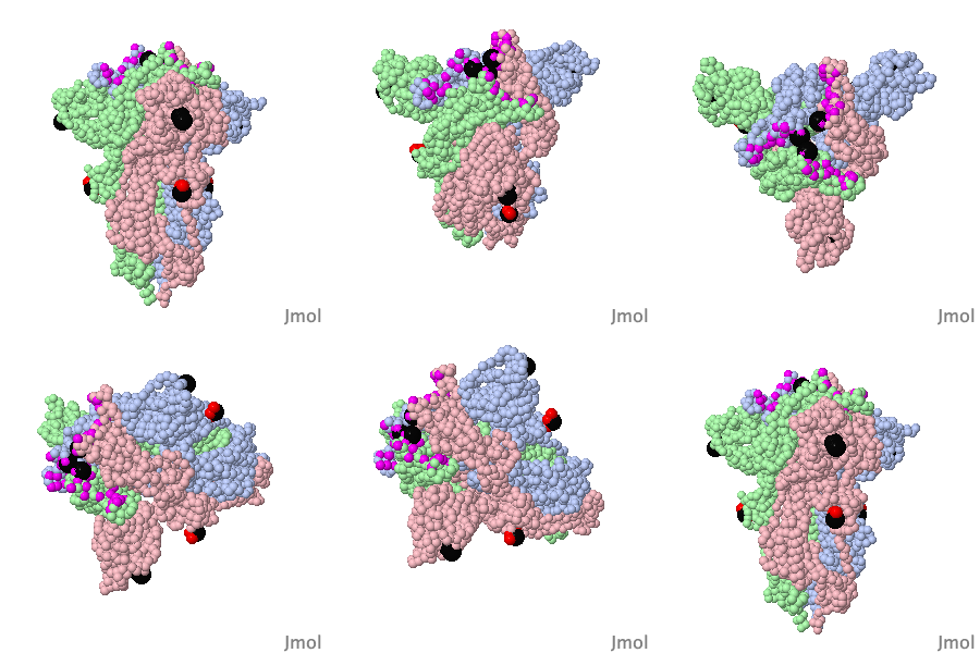
Coronavirus Spike Protein Priming

by Eric Martz
Coronavirus SARS-CoV-2 (responsible for COVID-19) has a spike protein on its surface, which enables it to infect host cells. Initially, proteases in the lungs clip the homo-trimeric spike protein at a unique sequence. This primes it, causing it to extend its receptor binding surface (shown in the above animation), optimizing binding to the host cell's ACE2 receptor (not shown). Next, spike protein initiates fusion of the virus and host cell membranes (not shown), enabling the virus RNA to enter the cell and initiate production of new virions. Knowledge of spike protein's molecular structure and function is crucial to developing effective therapies and vaccines.
>>> Visit this page >>>

About this image
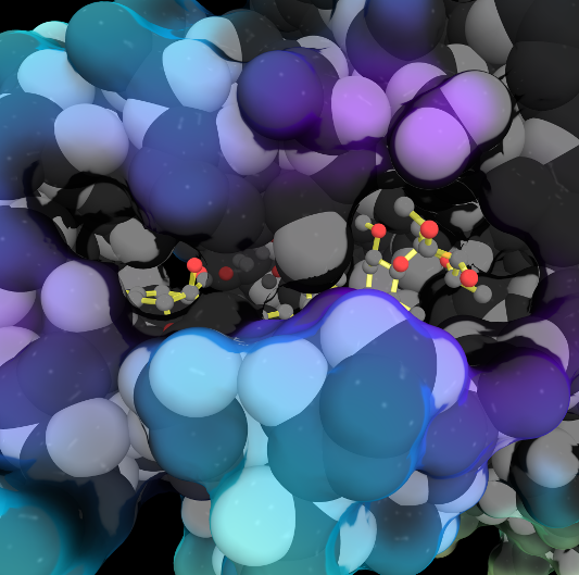
Molecular Sculpture

by Eric Martz
A historical review on sculptures and physical models of macromolecules.

>>> Visit this page >>>

About this image
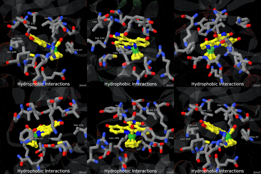
Structure of Anticancer Ruthenium Half-Sandwich Complex Bound to Glycogen Synthase Kinase 3ß

G Atilla-Gocumen, L Di Costanzo, E Meggers. J Biol Inorg Chem. 2010 doi: 10.1007/s00775-010-0699-x
A crystal structure of an organometallic half-sandwich ruthenium complex bound to glycogen synthase kinase 3ß (GSK-3ß) reveals that the inhibitor binds to the ATP binding site via an induced fit mechanism utilizing several hydrogen bonds and hydrophobic interactions. Importantly, the metal is not involved in any direct interaction with the protein kinase but fulfills a purely structural role.

>>> Visit this I3DC complement >>>

About this image
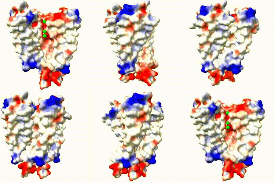
Polio is still here!
Polio vaccines have been available since the 1950s, but the challenges of vaccination in remote areas of Afghanistan and Pakistan have prevented worldwide eradication. In 2022, polio was found circulating in parts of New York State, USA. The polio virus has a small RNA genome enclosed in an icosahedral capsid composed of several proteins, shown cut in half. The structures of virus capsids can be explored using free FirstGlance in Jmol.

>>> Visit I3DC Interactive Visualizations >>>

How to add content to Proteopedia

Video Guides

Who knows ...

List of Art on Science pages

About Interactive 3D Complements - I3DCs

List of I3DCs

How to get an I3DC for your paper

Teaching strategies using Proteopedia

Examples of pages for teaching

How to add content to Proteopedia

About Contact Table of Contents Structure Index Help

Proteopedia Page Contributors and Editors (what is this?)

Joel L. Sussman, Jaime Prilusky

Personal tools