User:Estelle Blochouse/ Sandbox 1497
From Proteopedia
(Difference between revisions)
| Line 14: | Line 14: | ||
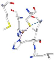
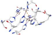
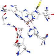
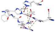
Multicopper oxidases are able to oxidise their substrates thanks to their particular structure. They own four copper ions (Cu1001, Cu1002, Cu1003 and C1004) dispatched between two important areas : the mononuclear copper center (Cu1001) and the trinuclear copper center (Cu1002, Cu1003 and Cu1004). | Multicopper oxidases are able to oxidise their substrates thanks to their particular structure. They own four copper ions (Cu1001, Cu1002, Cu1003 and C1004) dispatched between two important areas : the mononuclear copper center (Cu1001) and the trinuclear copper center (Cu1002, Cu1003 and Cu1004). | ||
| - | They accept an electron in the <scene name='pdbligand=CU:COPPER+(II)+ION'>mononoclear copper center</scene> and transfer it to the trinuclear copper center which binds to a molecule of dioxygen. The dioxygen receives four electrons from this transfer, therefore | + | They accept an electron in the <scene name='pdbligand=CU:COPPER+(II)+ION'>mononoclear copper center</scene> and transfer it to the trinuclear copper center which binds to a molecule of dioxygen. The dioxygen receives four electrons from this transfer, therefore it allows reduction of dioxygen into water. Dioxygen is transformed into two molecules of water.<ref>Bento I, Martins LO, Gato Lopes G, Arménia Carrondo M, Lindley PF, Dioxygen reduction by multi-copper oxidases; a structural perspective, November 2005</ref> Three differents copper centres exist that can be differentiated spectroscopically: Type 1 or blue (<scene name='pdbligand=CU:COPPER+(II)+ION'>mononoclear copper center</scene>)(Cu1001), type 2 or normal (Cu1004) and type 3 or coupled binuclear (Cu1002 and Cu1003).<ref>Messerschmidt A, Huber R, The blue oxidases, ascorbate oxidase, laccase and ceruloplasmin. Modelling and structural relationships, Eur. J. Biochem. 187, January 1990</ref><ref>Ouzounis C, Sander C, A structure-derived sequence pattern for the detection of type I copper binding domains in distantly related proteins, FEBS Lett. volume 279, February 1991</ref> |
== Mechanism == | == Mechanism == | ||
Revision as of 19:14, 11 January 2019
Multicopper Oxidase CueO (4e9s)
| |||||||||||
References
- ↑ EMBL-EBI, Family: Cu-oxidase (PF00394), Summary: Multicopper oxidase, http://pfam.xfam.org/family/Cu-oxidase
- ↑ UniProtKB
- ↑ Bento I, Martins LO, Gato Lopes G, Arménia Carrondo M, Lindley PF, Dioxygen reduction by multi-copper oxidases; a structural perspective, November 2005
- ↑ Messerschmidt A, Huber R, The blue oxidases, ascorbate oxidase, laccase and ceruloplasmin. Modelling and structural relationships, Eur. J. Biochem. 187, January 1990
- ↑ Ouzounis C, Sander C, A structure-derived sequence pattern for the detection of type I copper binding domains in distantly related proteins, FEBS Lett. volume 279, February 1991
- ↑ Hirofumi Komori, Ryosuke Sugiyama, Kunishige Kataoka, Kentaro Miyazaki, Yoshiki Higuchib, and Takeshi Sakurai, New insights into the catalytic active-site structure of multicopper oxidases, Biological Crystallography, 6 December 2013 doi:10.1107/S1399004713033051
- ↑ RCBS PDB
- ↑ RCBS PDB
- ↑ RCBS PDB
- ↑ RCBS PDB
- ↑ RCBS PDB
- ↑ RCBS PDB
- ↑ Kataoka K, Komori H, Ueki Y, Konno Y, Kamitaka Y, Kurose S, Tsujimura S, Higuchi Y, Kano K, Seo D, Sakurai T. Structure and function of the engineered multicopper oxidase CueO from Escherichia coli--deletion of the methionine-rich helical region covering the substrate-binding site. J Mol Biol. 2007 Oct 12;373(1):141-52. Epub 2007 Aug 2. PMID:17804014 doi:10.1016/j.jmb.2007.07.041
