We apologize for Proteopedia being slow to respond. For the past two years, a new implementation of Proteopedia has been being built. Soon, it will replace this 18-year old system. All existing content will be moved to the new system at a date that will be announced here.
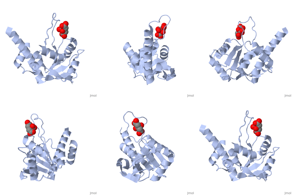
1dmg
From Proteopedia
(Difference between revisions)
| Line 3: | Line 3: | ||
<StructureSection load='1dmg' size='340' side='right'caption='[[1dmg]], [[Resolution|resolution]] 1.70Å' scene=''> | <StructureSection load='1dmg' size='340' side='right'caption='[[1dmg]], [[Resolution|resolution]] 1.70Å' scene=''> | ||
== Structural highlights == | == Structural highlights == | ||
| - | <table><tr><td colspan='2'>[[1dmg]] is a 1 chain structure. Full crystallographic information is available from [http://oca.weizmann.ac.il/oca-bin/ocashort?id=1DMG OCA]. For a <b>guided tour on the structure components</b> use [ | + | <table><tr><td colspan='2'>[[1dmg]] is a 1 chain structure. Full crystallographic information is available from [http://oca.weizmann.ac.il/oca-bin/ocashort?id=1DMG OCA]. For a <b>guided tour on the structure components</b> use [https://proteopedia.org/fgij/fg.htm?mol=1DMG FirstGlance]. <br> |
| - | </td></tr><tr id='ligand'><td class="sblockLbl"><b>[[Ligand|Ligands:]]</b></td><td class="sblockDat"><scene name='pdbligand=CIT:CITRIC+ACID'>CIT</scene></td></tr> | + | </td></tr><tr id='ligand'><td class="sblockLbl"><b>[[Ligand|Ligands:]]</b></td><td class="sblockDat" id="ligandDat"><scene name='pdbligand=CIT:CITRIC+ACID'>CIT</scene></td></tr> |
| - | <tr id='resources'><td class="sblockLbl"><b>Resources:</b></td><td class="sblockDat"><span class='plainlinks'>[ | + | <tr id='resources'><td class="sblockLbl"><b>Resources:</b></td><td class="sblockDat"><span class='plainlinks'>[https://proteopedia.org/fgij/fg.htm?mol=1dmg FirstGlance], [http://oca.weizmann.ac.il/oca-bin/ocaids?id=1dmg OCA], [https://pdbe.org/1dmg PDBe], [https://www.rcsb.org/pdb/explore.do?structureId=1dmg RCSB], [https://www.ebi.ac.uk/pdbsum/1dmg PDBsum], [https://prosat.h-its.org/prosat/prosatexe?pdbcode=1dmg ProSAT]</span></td></tr> |
</table> | </table> | ||
== Function == | == Function == | ||
| - | [[ | + | [[https://www.uniprot.org/uniprot/RL4_THEMA RL4_THEMA]] One of the primary rRNA binding proteins, this protein initially binds near the 5'-end of the 23S rRNA. It is important during the early stages of 50S assembly. It makes multiple contacts with different domains of the 23S rRNA in the assembled 50S subunit and ribosome (By similarity).[HAMAP-Rule:MF_01328_B] This protein only weakly controls expression of the E.coli S10 operon. It is incorporated into E.coli ribosomes, however it is not as firmly associated as the endogenous protein.[HAMAP-Rule:MF_01328_B] Forms part of the polypeptide exit tunnel (By similarity).[HAMAP-Rule:MF_01328_B] |
== Evolutionary Conservation == | == Evolutionary Conservation == | ||
[[Image:Consurf_key_small.gif|200px|right]] | [[Image:Consurf_key_small.gif|200px|right]] | ||
Revision as of 18:30, 10 March 2021
CRYSTAL STRUCTURE OF RIBOSOMAL PROTEIN L4
| |||||||||||
Categories: Large Structures | Huber, R | Wahl, M C | Worbs, M | Alpha-beta | Gene regulation | L4 | Ribosomal protein | Ribosome | Rna | S10 operon


