We apologize for Proteopedia being slow to respond. For the past two years, a new implementation of Proteopedia has been being built. Soon, it will replace this 18-year old system. All existing content will be moved to the new system at a date that will be announced here.
5-hydroxytryptamine receptor
From Proteopedia
(Difference between revisions)
| Line 17: | Line 17: | ||
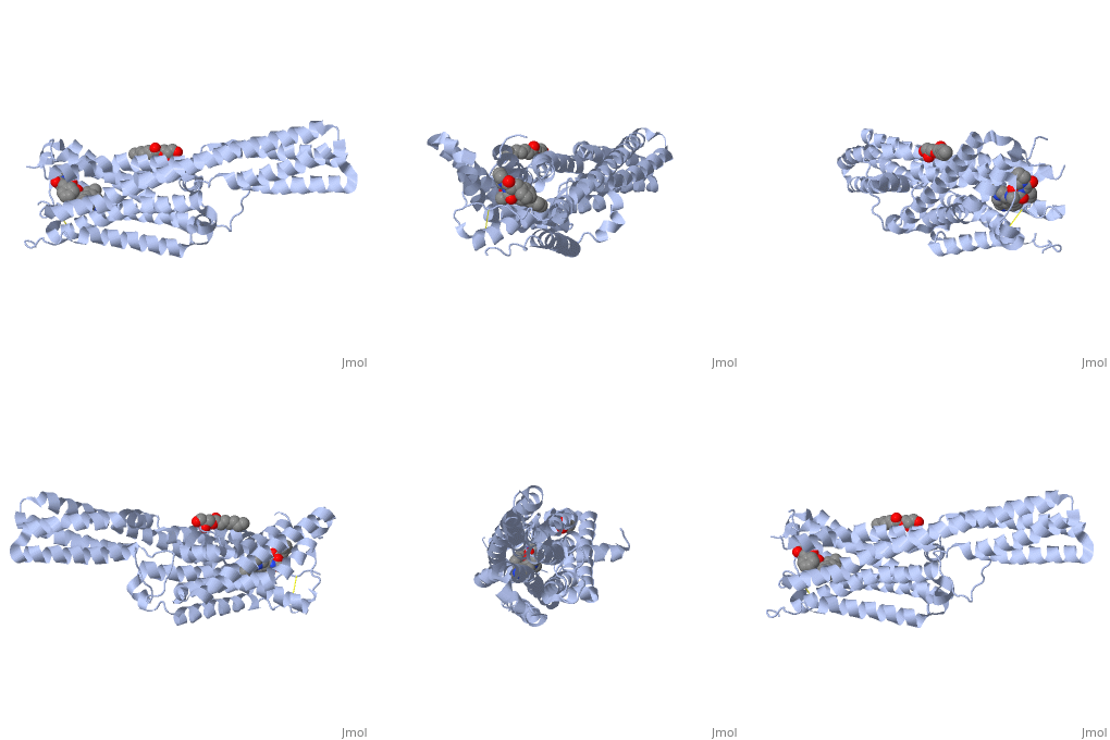
<scene name='71/716548/5-ht3_receptor/1'>The 5-HT3 receptor</scene> is a pentameric cation-selective ion channel and plays a role in neuronal excitation to release neurotransmitters from the postsynaptic neuron. Opening of the cation channel causes an influx of sodium and calcium through the receptor pore leading to a membrane depolarization. Five receptor subunits, A to E, have been found in humans but only subunits A and B have been found in rodents. When experimentally expressed in a host, the 5-HT3 receptor is comprised of either A or AB subunits which can result in a homopentameric receptor or a heteropentameric receptor respectively. The A and B subunits are found throughout the brain in areas such as the hippocampus and amygdala. 5-HT3 is a transmembrane channel that is stimulated to open state by the interaction of the receptor with serotonin in the extracellular space.<ref>Hassaine G,Cedric D, Luigino G, Romain W, Menno BT, Ruud H, Alexandra G, Henning S, Takashi T, Aline D, Christophe M, Xiao-Dan L, Frederic P, Horst V, Hugues N. ''X-ray Structure of the Mouse Serotonin 5-HT3 Receptor. Nature 512.7514 (2014): 276-81.[http://www.nature.com/nature/journal/v512/n7514/full/nature13552.html DOI:10.1038/nature13552]</ref> The binding site is comprised of six loops from two adjacent subunits in the extracellular N-terminal domain. Loops A, B and C form the principal subunit and contain the <scene name='71/716548/5-ht3/1'>important side chains</scene> N128, W183 and Y234. Loops D, E and F form the complementary subunit of the binding site and contain the important side chains W90, Y143 and W195. The transmembrane region is comprised of multiple alpha helical structures and mediates ion flow and ion specificity.<ref name = two> Thompson AJ, Lummis SCR. 5-HT3 Receptors. Curr Pharm Des. 2006; 12(28): 3615–3630. PMID:2664614 [http://www.ncbi.nlm.nih.gov/pmc/articles/PMC2664614/]</ref><br /> | <scene name='71/716548/5-ht3_receptor/1'>The 5-HT3 receptor</scene> is a pentameric cation-selective ion channel and plays a role in neuronal excitation to release neurotransmitters from the postsynaptic neuron. Opening of the cation channel causes an influx of sodium and calcium through the receptor pore leading to a membrane depolarization. Five receptor subunits, A to E, have been found in humans but only subunits A and B have been found in rodents. When experimentally expressed in a host, the 5-HT3 receptor is comprised of either A or AB subunits which can result in a homopentameric receptor or a heteropentameric receptor respectively. The A and B subunits are found throughout the brain in areas such as the hippocampus and amygdala. 5-HT3 is a transmembrane channel that is stimulated to open state by the interaction of the receptor with serotonin in the extracellular space.<ref>Hassaine G,Cedric D, Luigino G, Romain W, Menno BT, Ruud H, Alexandra G, Henning S, Takashi T, Aline D, Christophe M, Xiao-Dan L, Frederic P, Horst V, Hugues N. ''X-ray Structure of the Mouse Serotonin 5-HT3 Receptor. Nature 512.7514 (2014): 276-81.[http://www.nature.com/nature/journal/v512/n7514/full/nature13552.html DOI:10.1038/nature13552]</ref> The binding site is comprised of six loops from two adjacent subunits in the extracellular N-terminal domain. Loops A, B and C form the principal subunit and contain the <scene name='71/716548/5-ht3/1'>important side chains</scene> N128, W183 and Y234. Loops D, E and F form the complementary subunit of the binding site and contain the important side chains W90, Y143 and W195. The transmembrane region is comprised of multiple alpha helical structures and mediates ion flow and ion specificity.<ref name = two> Thompson AJ, Lummis SCR. 5-HT3 Receptors. Curr Pharm Des. 2006; 12(28): 3615–3630. PMID:2664614 [http://www.ncbi.nlm.nih.gov/pmc/articles/PMC2664614/]</ref><br /> | ||
For more details see [[5-ht3a receptor]] and [[Ion channels]]. | For more details see [[5-ht3a receptor]] and [[Ion channels]]. | ||
| + | |||
| + | === The extracellular subunit interface of the 5-HT3 receptors: a computational alanine scanning mutagenesis study === | ||
| + | <big>Francesca De Rienzo, Arménio J. Moura Barbosa, Marta A.S. Perez, Pedro A. Fernandes, Maria J. Ramos, Maria Cristina Menziani</big> <ref>DOI 10.1080/07391102.2012.680029</ref> | ||
| + | <hr/> | ||
| + | <b>Molecular Tour</b><br> | ||
| + | The serotonin type-3 receptor (5-HT3-R) is a cation selective transmembrane protein channel that belongs to the Cys–loop Ligand-Gated Ion Channel (LGIC) superfamily (http://www.ebi.ac.uk/compneur-srv/LGICdb/LGICdb.php), which also includes receptors for nicotinic acetylcholine (<scene name='Journal:JBSD:16/Cv/2'>nAChR</scene>, PDB code [[2bg9]]), γ-aminobutyric acid and glycine. 5-HT3-R is involved in signal transmission in the central and peripheral nervous system and its malfunctioning leads to neurodegenerative and psychiatric diseases, therefore it is an important target for drug design research. A few drugs active against 5-HT3-R are already on the market, such as, for example, palonosetron (http://en.wikipedia.org/wiki/Palonosetron) and granisetron (http://en.wikipedia.org/wiki/Granisetron). | ||
| + | The 5-HT3R is made of five monomers assembled in a <scene name='Journal:JBSD:16/Cv/4'>pseudo-symmetric pentameric shape</scene> to form an ion channel permeable to small ions (Na+, K+); each subunit contains three domains: an <scene name='Journal:JBSD:16/Cv/3'>intracellular portion, a transmembrane domain and an extracellular region</scene> (shown on the example of nAChR, [[2bg9]]). To date, five different 5-HT3-R subunits have been identified, the 5-HT3 A, B, C, D and E; however, only subunits A and B have been extensively characterised experimentally. The <scene name='Journal:JBSD:16/Cv/6'>ligand binding site</scene> of nAChR is located at the extracellular region, at the interface between two monomers (α-γ and α-δ; 2 identical α monomers, chains A and D, are colored in same color - lavender), called the principal and the complementary subunits. | ||
| + | The 3D structure of 5-HT3-R has not been experimentally solved yet; however, it has been obtained computationally by means of homology modelling techniques. (http://salilab.org/modeller/) | ||
| + | Thus, the <scene name='Journal:JBSD:16/Cv/7'>extracellular region of the 5HT3 subunits A and B</scene> are modelled by homology with the 3D structure of the nAChR subunit A ([[2bg9]]-A) and are used to assemble receptor structures as pseudo-symmetric pentamers made either of <scene name='Journal:JBSD:16/Cv/9'>five identical subunits A (homomeric 5-HT3A-R)</scene> or of <scene name='Journal:JBSD:16/Cv/10'>both subunits A and B (heteromeric 5-HT3A/B-R in the BBABA arrangement)</scene> in a still debated arrangement.<ref>PMID:20724042 </ref> Subunits <font color='magenta'><b>A</b></font> and <font color='red'><b>B</b></font> are colored in <font color='magenta'><b>magenta</b></font> and <font color='red'><b>red</b></font>, respectively. | ||
| + | A complete characterization of the extracellular moiety of the <scene name='Journal:JBSD:16/Cv/15'>dimer interface of the 5-HT3-R</scene> (AA dimer is shown, <span style="color:cyan;background-color:black;font-weight:bold;">principal subunit is colored in cyan</span> and <font color='blue'><b>complementary is in blue</b></font>, is obtained by the Computational Alanine Scanning Mutagenesis (CASM) approach <ref>PMID:17195156</ref>, which simulates the substitution, one by one, of all the amino acid residues at the subunit-subunit interfaces with an Ala, thus to assess the interface binding contribution of single residue side-chains. The <scene name='Journal:JBSD:16/Cv/16'>most relevant residues for interface stabilization</scene> are classified as “hot spots” that stabilize the interface by more than 4 kcal/mol and “warm spots” that contribute to interface stabilization by more than 2 kcal/mol. <scene name='Journal:JBSD:16/Cv/17'>Click here to see also the interface of complementary subunit.</scene> Interface residues are shown in spacefill representation, <font color='red'><b>hot spot residues are colored in red</b></font> and <span style="color:orange;background-color:black;font-weight:bold;">warm spots residues are are in orange</span>. | ||
| + | From this analysis the <scene name='Journal:JBSD:16/Cv/18'>important aromatic cluster</scene> located at the interface core and formed by residues W178 (principal subunit), Y68, Y83, W85 and Y148 (complementary subunit) is highlighted.<ref>DOI:10.1080/07391102.2012.680029</ref> In addition, two important groups of interface residues are probably involved in the coupling of <scene name='Journal:JBSD:16/Cv1/6'>agonist</scene> and <scene name='Journal:JBSD:16/Cv1/10'>antagonist</scene> binding to channel activation/inactivation: W116-H180-L179-W178-E124-F125 (principal subunit) and Y136-Y138-Y148-W85-(P150) (complementary subunit), where W178 and Y148 appear to be critical residues for the binding/activation mechanism. Finally, the <scene name='Journal:JBSD:16/Cv1/8'>comparison of the AA interface with the BB interface</scene> (<span style="color:cyan;background-color:black;font-weight:bold;">principal subunit of AA is colored in cyan</span>, <font color='darkmagenta'><b>principal subunit BB is colored in darkmagenta</b></font>, <font color='blue'><b>complementary subunit AA is in blue</b></font> and <font color='magenta'><b>complementary subunit BB is in magenta</b></font>) shows differences which could explain the reasons why the homopentamer 5-HT3B-R, if expressed, is not functional (see also image below).<ref>DOI: 10.1039/C2CP41028A</ref> | ||
| + | |||
| + | [[Image:Fig 7.png|left|400px|thumb|]] | ||
== 5HT-2B receptor agonists: Lysergic Acid Diethylamide (LSD)== | == 5HT-2B receptor agonists: Lysergic Acid Diethylamide (LSD)== | ||
Revision as of 14:36, 12 November 2019
| |||||||||||
References
- ↑ Goodsell D. Serotonin Receptor. RCSB PDB-101 (2013) DOI: 10.2210/rcsb_pdb/mom_2013_8
- ↑ 2.0 2.1 2.2 2.3 Wang C, Jiang Y, Ma J, Wu H, Wacker D, Katritch V, Han GW, Liu W, Huang XP, Vardy E, McCorvy JD, Gao X, Zhou EZ, Melcher K, Zhang C, Bai F, Yang H, Yang L, Jiang H, Roth BL, Cherezov V, Stevens RC, Xu HE. Structural Basis for Molecular Recognition at Serotonin Receptors. Science. 2013 May 3; 340(6132): 610–614. PMID:3644373 [1]
- ↑ Wiebke J, Schymura Y, Novoyatleva T, Kojonazarov B, Boehm M, Wietelmann A, Luitel H, Murmann K, Krompiec DR, Tretyn A, Pullamsetti SS, Weissmann N, Seeger W, Ghofrani HA, Schermuly RT. 5-HT2B Receptor Antagonists Inhibit Fibrosis and Protect from RV Heart Failure. Biomed Res Int. 2015; 2015: 438403. PMID:4312574 [2]
- ↑ Nebigil, Etienne, Schaerlinger, Hickel, Launay, Maroteaux. Developmentally Regulated Serotonin 5-HT2B Receptors. DOI: 10.1016/S0736-5748(01)00022-3
- ↑ Berumen LC, Rodriguez A, Miledi R, Gracia-Alcocer G. Serotonin Receptors in Hippocampus. ScientificWorldJournal. 2012;2012:823493. Epub 2012 May 2. PMID:3353568 [3]
- ↑ Millan MJ. Serotonin 5-HT2C receptors as a target for the treatment of depressive and anxious states: focus on novel therapeutic strategies. Therapie. 2005 Sep-Oct;60(5):441-60. PMID:16433010
- ↑ Ge T, Zhang Z, Lv J, Song Y, Fan J, Liu W, Wang X, Hall FS, Li B, Cui R. The role of 5-HT2c receptor on corticosterone-mediated food intake. J Biochem Mol Toxicol. 2017 Jun;31(6). doi: 10.1002/jbt.21890. Epub 2017 Feb 10. PMID:28186389 doi:http://dx.doi.org/10.1002/jbt.21890
- ↑ Hassaine G,Cedric D, Luigino G, Romain W, Menno BT, Ruud H, Alexandra G, Henning S, Takashi T, Aline D, Christophe M, Xiao-Dan L, Frederic P, Horst V, Hugues N. X-ray Structure of the Mouse Serotonin 5-HT3 Receptor. Nature 512.7514 (2014): 276-81.DOI:10.1038/nature13552
- ↑ 9.0 9.1 9.2 Thompson AJ, Lummis SCR. 5-HT3 Receptors. Curr Pharm Des. 2006; 12(28): 3615–3630. PMID:2664614 [4]
- ↑ De Rienzo F, Moura Barbosa AJ, Perez MA, Fernandes PA, Ramos MJ, Menziani MC. The extracellular subunit interface of the 5-HT(3) receptors: a computational alanine scanning mutagenesis study. J Biomol Struct Dyn. 2012 Jul;30(3):280-98. Epub 2012 Jun 12. PMID:22694192 doi:10.1080/07391102.2012.680029
- ↑ Moura Barbosa AJ, De Rienzo F, Ramos MJ, Menziani MC. Computational analysis of ligand recognition sites of homo- and heteropentameric 5-HT3 receptors. Eur J Med Chem. 2010 Nov;45(11):4746-60. Epub 2010 Jul 27. PMID:20724042 doi:10.1016/j.ejmech.2010.07.039
- ↑ Moreira IS, Fernandes PA, Ramos MJ. Computational alanine scanning mutagenesis--an improved methodological approach. J Comput Chem. 2007 Feb;28(3):644-54. PMID:17195156 doi:10.1002/jcc.20566
- ↑ De Rienzo F, Moura Barbosa AJ, Perez MA, Fernandes PA, Ramos MJ, Menziani MC. The extracellular subunit interface of the 5-HT(3) receptors: a computational alanine scanning mutagenesis study. J Biomol Struct Dyn. 2012 Jul;30(3):280-98. Epub 2012 Jun 12. PMID:22694192 doi:10.1080/07391102.2012.680029
- ↑ De Rienzo F, Del Cadia M, Menziani MC. A first step towards the understanding of the 5-HT(3) receptor subunit heterogeneity from a computational point of view. Phys Chem Chem Phys. 2012 Sep 28;14(36):12625-36. Epub 2012 Aug 9. PMID:22880201 doi:10.1039/c2cp41028a
- ↑ Maksay G, Zsolt B, Miklós S. Binding Interactions of Antagonists with 5‐Hydroxytryptamine 3A Receptor Models. Journal of Receptors and Signal Transduction 23.2-3 (2003): 255-70. DOI:10.1081/RRS-120025568
- ↑ Brunton LL, Lazo JS, Parker KL. (2006). Goddman & Gilman's The Pharmacological Basis of Therapeutics. New York: McGraw-Hill. pp. 1000–3. ISBN 978-0-07-142280-2.
Proteopedia Page Contributors and Editors (what is this?)
Michal Harel, Alexander Berchansky, Joel L. Sussman, Zachary Falk, Brittany Spence, Matthew P Cabrera, Jaime Prilusky


