We apologize for Proteopedia being slow to respond. For the past two years, a new implementation of Proteopedia has been being built. Soon, it will replace this 18-year old system. All existing content will be moved to the new system at a date that will be announced here.
Sandbox Reserved 1568
From Proteopedia
(Difference between revisions)
| Line 17: | Line 17: | ||
LsdA appears, when crystallized, as two LsdA protomers in one asymmetric unit as a dimer. The <scene name='82/823092/Secondary_and_tertiary_struct/1'>secondary and tertiary structure</scene> of the protomer consists of α-helices (purple) and ß-sheets (blue). The ß-sheets are arranged in a <scene name='82/823092/Seven-bladed_beta_propeller/1'>seven-bladed ß-propeller</scene>, typical of the carotenoid cleavage oxygenases. | LsdA appears, when crystallized, as two LsdA protomers in one asymmetric unit as a dimer. The <scene name='82/823092/Secondary_and_tertiary_struct/1'>secondary and tertiary structure</scene> of the protomer consists of α-helices (purple) and ß-sheets (blue). The ß-sheets are arranged in a <scene name='82/823092/Seven-bladed_beta_propeller/1'>seven-bladed ß-propeller</scene>, typical of the carotenoid cleavage oxygenases. | ||
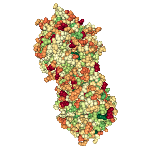
| + | When you look at the <scene name='82/823092/Spacefill_lsda/1'>spacefill view</scene> of the protein dimer you see that the binding pocket accessibility is very restrictive. | ||
| + | [[Image:spacefill hydrophobicity.png]][[Image:ligand hydrophobicity]] | ||
| + | Hydrophobicity-focused view of the protein. | ||
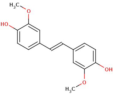
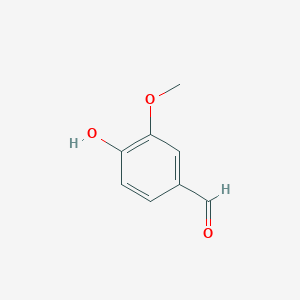
The <scene name='82/823092/Catalytic_triad/2'>catalytic triad</scene> of the binding site consists of Phe59, Tyr101, and Lys134 that contact the 4-hydroxyphenyl portion of the substrate. | The <scene name='82/823092/Catalytic_triad/2'>catalytic triad</scene> of the binding site consists of Phe59, Tyr101, and Lys134 that contact the 4-hydroxyphenyl portion of the substrate. | ||
Revision as of 00:47, 30 November 2019
| This Sandbox is Reserved from Aug 26 through Dec 12, 2019 for use in the course CHEM 351 Biochemistry taught by Bonnie_Hall at the Grand View University, Des Moines, USA. This reservation includes Sandbox Reserved 1556 through Sandbox Reserved 1575. |
To get started:
More help: Help:Editing |
Lignostilbene-α,ß-dioxygenase A structural features and important functional residues
| |||||||||||
References
- ↑ Hanson, R. M., Prilusky, J., Renjian, Z., Nakane, T. and Sussman, J. L. (2013), JSmol and the Next-Generation Web-Based Representation of 3D Molecular Structure as Applied to Proteopedia. Isr. J. Chem., 53:207-216. doi:http://dx.doi.org/10.1002/ijch.201300024
- ↑ Herraez A. Biomolecules in the computer: Jmol to the rescue. Biochem Mol Biol Educ. 2006 Jul;34(4):255-61. doi: 10.1002/bmb.2006.494034042644. PMID:21638687 doi:10.1002/bmb.2006.494034042644
- ↑ Kuatsjah E, Verstraete MM, Kobylarz MJ, Liu AKN, Murphy MEP, Eltis LD. Identification of functionally important residues and structural features in a bacterial lignostilbene dioxygenase. J Biol Chem. 2019 Jul 10. pii: RA119.009428. doi: 10.1074/jbc.RA119.009428. PMID:31292192 doi:http://dx.doi.org/10.1074/jbc.RA119.009428


