Sandbox Reserved 1600
From Proteopedia
(Difference between revisions)
| Line 14: | Line 14: | ||
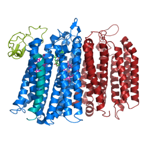
The overall structure contains 19 transmembrane helices that are arranged in a nearly oval shape (Fig 1.). The protein contains two structurally similar subunits each containing nine helices (blue and red) and one smaller subunit, CydX, with one transmembrane helix. The subunits are interacting using hydrophobic residues and symmetry at the interfaces. The CydX subunit, whose function is not currently known, is positioned in the same way as CydS, which is found in E. coli bd oxidase. Due to its similar structure and position, it has been hypothesized to potentially stabilize heme b558 during potential structural rearrangements of the Q loop upon binding and oxidation of quinol. The Q loop is shown in lime green, and is a hydrophilic region above Cyd A. The lack of hydrogen bonding in this hydrophobic protein allows the protein to be flexible and go through a large conformational change for reduction of dioxygen. | The overall structure contains 19 transmembrane helices that are arranged in a nearly oval shape (Fig 1.). The protein contains two structurally similar subunits each containing nine helices (blue and red) and one smaller subunit, CydX, with one transmembrane helix. The subunits are interacting using hydrophobic residues and symmetry at the interfaces. The CydX subunit, whose function is not currently known, is positioned in the same way as CydS, which is found in E. coli bd oxidase. Due to its similar structure and position, it has been hypothesized to potentially stabilize heme b558 during potential structural rearrangements of the Q loop upon binding and oxidation of quinol. The Q loop is shown in lime green, and is a hydrophilic region above Cyd A. The lack of hydrogen bonding in this hydrophobic protein allows the protein to be flexible and go through a large conformational change for reduction of dioxygen. | ||
| - | == | + | ==Active Site== |
| - | ( | + | |
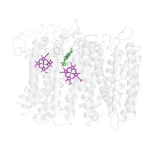
| + | The active site for Bd Oxidase in ''Geobacillus thermodenitrificans'' is located in subunit Cyd A. The site consists of three iron hemes: Heme B558, Heme B595, and Heme D shown in Fig.2. [[Image:Hemes.jpg|300 px|right|thumb|Figure 2. The active site of Bd Oxidase. Heme D is shown with green carbons. Heme B558 and Heme B595 are shown on the left and right respectively with pink carbons.]] The three hemes are held together in a rigid triangular arrangement due to van der wals interactions. The length between each heme's central iron is relatively constant which serves to shuttle protons and electrons from one heme to another efficiently. It is suggested that Heme B558 acts as an electron acceptor to the extracellular side and Heme B559 acts as a proton acceptor on the intracellular side. It is then proposed that both heme B558 and B595 shuttle their respective ions directly to Heme D based on this being the shortest pathway (reference). Heme D is then suggested to be the oxygen binding site due to proximity and orientation to the exterior surface of the protein. | ||
==Potential Oxygen Entry Site== | ==Potential Oxygen Entry Site== | ||
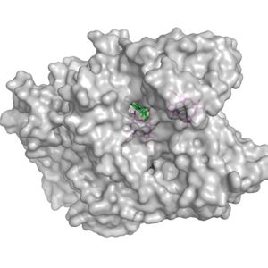
| - | [[Image:Potential_oxygen_entry_site.jpg|300 px|right|thumb|Figure | + | [[Image:Potential_oxygen_entry_site.jpg|300 px|right|thumb|Figure 3. Surface of the potential oxygen entry site; Heme D shown in green]] |
| - | Heme D is the hypothesized spot for the oxygen to enter the protein. Heme D is directly connected to the protein surface on CydA and contains a solvent accessible substrate channel (Fig | + | Heme D is the hypothesized spot for the oxygen to enter the protein. Heme D is directly connected to the protein surface on CydA and contains a solvent accessible substrate channel (Fig 3.) |
==Electron Source== | ==Electron Source== | ||
Revision as of 15:19, 23 March 2020
bd oxidase; Geobacillus thermodenitrificans
| |||||||||||


