We apologize for Proteopedia being slow to respond. For the past two years, a new implementation of Proteopedia has been being built. Soon, it will replace this 18-year old system. All existing content will be moved to the new system at a date that will be announced here.
Sandbox Reserved 1607
From Proteopedia
(Difference between revisions)
| Line 10: | Line 10: | ||
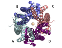
The selectivity pore is an integral part of the protein. This pore contains a group of glutamate with oxygen facing inward forming a carboxylate ring through which calcium enters. This negative carboxylate ring does a good job of pulling the positive calcium into the selectivity pore at the top of the protein. | The selectivity pore is an integral part of the protein. This pore contains a group of glutamate with oxygen facing inward forming a carboxylate ring through which calcium enters. This negative carboxylate ring does a good job of pulling the positive calcium into the selectivity pore at the top of the protein. | ||
| - | [https://en.wikipedia.org/wiki/Cryogenic_electron_microscopy Cryogenic electron microscopy] (Cryo-EM) was instrumental in outlining the complete structure of this protein. <ref>DOI 10.1038/s41580-018-0052-8</ref> | + | [https://en.wikipedia.org/wiki/Cryogenic_electron_microscopy Cryogenic electron microscopy] (Cryo-EM) was instrumental in outlining the complete structure of this protein. <ref name="Giorgi C">DOI 10.1038/s41580-018-0052-8</ref> |
== Structural highlights and mechanism == | == Structural highlights and mechanism == | ||
| Line 34: | Line 34: | ||
===Types 1 and 2 Diabetes=== | ===Types 1 and 2 Diabetes=== | ||
| - | Pancreatic [https://en.wikipedia.org/wiki/Beta_cell beta cells] circulate insulin through the body. Glucose initiates signals allowing these cells to break down sugar and release insulin, which is all stimulated by mitochondrial energy metabolism. Calcium homeostasis plays a fundamental role in ATP production supplying energy to this process. Defects in homeostasis of calcium like chronic calcium depletion, caused by leaky [https://en.wikipedia.org/wiki/Ryanodine_receptor Ryanodine receptors], causes types [https://en.wikipedia.org/wiki/Type_1_diabetes 1] and [https://en.wikipedia.org/wiki/Type_2_diabetes 2] diabetes through failure of this mechanism. Treatment involves targeting the MCU and MICU1 to open the calcium channel and allow more uptake of calcium ions into the mitochondria. <ref | + | Pancreatic [https://en.wikipedia.org/wiki/Beta_cell beta cells] circulate insulin through the body. Glucose initiates signals allowing these cells to break down sugar and release insulin, which is all stimulated by mitochondrial energy metabolism. Calcium homeostasis plays a fundamental role in ATP production supplying energy to this process. Defects in homeostasis of calcium like chronic calcium depletion, caused by leaky [https://en.wikipedia.org/wiki/Ryanodine_receptor Ryanodine receptors], causes types [https://en.wikipedia.org/wiki/Type_1_diabetes 1] and [https://en.wikipedia.org/wiki/Type_2_diabetes 2] diabetes through failure of this mechanism. Treatment involves targeting the MCU and MICU1 to open the calcium channel and allow more uptake of calcium ions into the mitochondria. <ref name="Giorgi C" /> |
===Heart Failure=== | ===Heart Failure=== | ||
| - | Calcium overload in the mitochondria of cardiac cells lead to [https://en.wikipedia.org/wiki/Apoptosis apoptotic] cardiac cell death. Calcium governs [https://en.wikipedia.org/wiki/Cardiac_excitation-contraction_coupling excitation contraction coupling] (EC coupling) of the cardiac muscles, which creates the ATP needed to power the contraction during heart beats. The increase in mitochondrial Ca2+ concentration is essential for the functioning of this muscle contraction. Mitochondrial Ca2+ overload, though, leads to necrotic cardiac cell death and can be targeted with regulation of the MCU. An example of treatment would be with the use of Ru360 (Figure 3) to inhibit the uptake of Ca2+ ions into the mitochondria. <ref | + | Calcium overload in the mitochondria of cardiac cells lead to [https://en.wikipedia.org/wiki/Apoptosis apoptotic] cardiac cell death. Calcium governs [https://en.wikipedia.org/wiki/Cardiac_excitation-contraction_coupling excitation contraction coupling] (EC coupling) of the cardiac muscles, which creates the ATP needed to power the contraction during heart beats. The increase in mitochondrial Ca2+ concentration is essential for the functioning of this muscle contraction. Mitochondrial Ca2+ overload, though, leads to necrotic cardiac cell death and can be targeted with regulation of the MCU. An example of treatment would be with the use of Ru360 (Figure 3) to inhibit the uptake of Ca2+ ions into the mitochondria. <ref name="Giorgi C" /> |
== Student Contributors == | == Student Contributors == | ||
Revision as of 04:41, 7 April 2020
Mitochondrial Calcium Uniporter
| |||||||||||
References
- ↑ Hanson, R. M., Prilusky, J., Renjian, Z., Nakane, T. and Sussman, J. L. (2013), JSmol and the Next-Generation Web-Based Representation of 3D Molecular Structure as Applied to Proteopedia. Isr. J. Chem., 53:207-216. doi:http://dx.doi.org/10.1002/ijch.201300024
- ↑ Herraez A. Biomolecules in the computer: Jmol to the rescue. Biochem Mol Biol Educ. 2006 Jul;34(4):255-61. doi: 10.1002/bmb.2006.494034042644. PMID:21638687 doi:10.1002/bmb.2006.494034042644
- ↑ 3.0 3.1 3.2 Giorgi C, Marchi S, Pinton P. The machineries, regulation and cellular functions of mitochondrial calcium. Nat Rev Mol Cell Biol. 2018 Nov;19(11):713-730. doi: 10.1038/s41580-018-0052-8. PMID:30143745 doi:http://dx.doi.org/10.1038/s41580-018-0052-8
- ↑ 4.0 4.1 4.2 4.3 4.4 Fan C, Fan M, Orlando BJ, Fastman NM, Zhang J, Xu Y, Chambers MG, Xu X, Perry K, Liao M, Feng L. X-ray and cryo-EM structures of the mitochondrial calcium uniporter. Nature. 2018 Jul 11. pii: 10.1038/s41586-018-0330-9. doi:, 10.1038/s41586-018-0330-9. PMID:29995856 doi:http://dx.doi.org/10.1038/s41586-018-0330-9
- ↑ Yoo J, Wu M, Yin Y, Herzik MA Jr, Lander GC, Lee SY. Cryo-EM structure of a mitochondrial calcium uniporter. Science. 2018 Jun 28. pii: science.aar4056. doi: 10.1126/science.aar4056. PMID:29954988 doi:http://dx.doi.org/10.1126/science.aar4056


