We apologize for Proteopedia being slow to respond. For the past two years, a new implementation of Proteopedia has been being built. Soon, it will replace this 18-year old system. All existing content will be moved to the new system at a date that will be announced here.
Sandbox Reserved 1627
From Proteopedia
(Difference between revisions)
| Line 22: | Line 22: | ||
===Conformation Change=== | ===Conformation Change=== | ||
| - | + | When the receptor is in an <scene name='83/832953/Inactive_insulin_receptor/3'>inverted V</scene> shape, the FnIII-3 domains are separated by about 120Å. This distance prevents the initiation of autophosphorylation and downstream signaling by the tyrosine kinase domains on the intracellular side of the receptor. Upon the binding of insulin to three binding sites, 1, 1', and either 2 or 2', the conformation change will begin and bring the FnIII-3 domains within 40Å of each other to induce the <scene name='83/832953/Ir_dimer_t_state/3'>T shape</scene> conformation. <ref> DOI 10.1038/s41467-018-06826-6</ref> <ref name="Uchikawa" /> The T shape conformation is well observed in the alpha subunit. It is horizontally composed of L1, CR (including the α-CT chain), and L2 domains and vertically composed of the FnIII-1, 2, and 3 domains. The insulin receptor's structural [http://en.wikipedia.org/wiki/Conformational_change conformation change] is what allows it to go from the inactive state to the active state in order to facilitate the autophosphorylation of the tyrosine kinase domain. | |
===Binding interactions=== | ===Binding interactions=== | ||
| - | [[Image:Binding site with AA labeled.png|thumb|right|270px|Figure 3: Subunit interactions between the insulin receptor CT-alpha helix (light blue) and insulin (magenta) in one of the binding sites. [http://www.rcsb.org/structure/6sof PDB 6SOF]]] | ||
| - | |||
| - | The actual site of insulin binding occurs at the <scene name='83/832953/Alpha_c_helix/5'>α-CT chain</scene> of one of the sites discussed next and is stabilized by the L1 and L2 domains. | ||
| - | |||
| - | "Cross linking" | ||
| - | |||
| - | A tripartite interaction occurs between the alpha-CT chain and the FnIII-1 domain region during the conformational change. This interaction involves the following residues: <scene name='83/832953/Alpha_ct_and_fniii-1/7'>ASP496, ARG498, and ASP499 on the FnIII-1 domain</scene> and the <scene name='83/832953/Alpha_ct_and_fniii-1/9'>LYS703, GLU706, and ASP707 on the alpha-CT domain</scene>. This duo then interacts with the leucine rich region, L1, that exists on the opposing protomer of the dimer. The tripartite interaction between the alpha-CT chain and FnIII-1 domain on one dimer and the L1 region on the other dimer is important because it allows for a strong and stable interaction between two dimers of the insulin receptor that maintains the T-shape activation state for the rest of the downstream signaling to occur. | ||
| - | |||
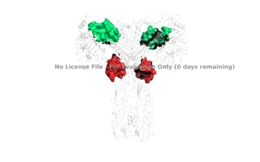
[[Image:4 sites highlighted.png|thumb|right|260px|Figure 4: The presence of all four potential binding sides on the active insulin receptor: sites 1 and 1' (green) and sites 2, and 2'(red). [http://www.rcsb.org/structure/6sof PDB 6SOF]]] | [[Image:4 sites highlighted.png|thumb|right|260px|Figure 4: The presence of all four potential binding sides on the active insulin receptor: sites 1 and 1' (green) and sites 2, and 2'(red). [http://www.rcsb.org/structure/6sof PDB 6SOF]]] | ||
| - | + | A tripartite interaction occurs between three critical parts of the alpha subunits of the insulin receptor. One one subunit, the α-CT chain and the FnIII-1 domain region become in close proximity during the conformational change of the insulin receptor. This interaction involves the following residues: <scene name='83/832953/Alpha_ct_and_fniii-1/7'>ASP496, ARG498, and ASP499 on the FnIII-1 domain</scene> and the <scene name='83/832953/Alpha_ct_and_fniii-1/9'>LYS703, GLU706, and ASP707 on the α-CT domain</scene>. This duo then interacts with the leucine rich region, L1, that exists on the opposing alpha subunit of the dimer. The fact that the two alpha subunits are interacting displays a "cross linking" scenario where the domains of the heterodimer can intertwine with each other. The tripartite interaction between the α-CT chain, FnIII-1 domain, and the L1 region is important because it allows for a strong and stable interaction between two subunits of the insulin receptor that maintains the T-shape activation state for the rest of the downstream signaling to occur. | |
| - | Although interactions at all four binding sites are highly hydrophobic, the ligand binding interactions at sites 1 and 1' are different than at sites 2 and 2'. Sites 1 and 1' | + | For insulin binding to induce the activation of the receptor and change its conformation to the active T state, binding at sites 1 and 1', as well as one insulin to either binding site 2 or 2', is required. <ref> DOI 10.7554/eLife.48630 </ref>. Although interactions at all four binding sites are highly hydrophobic, the ligand binding interactions at sites 1 and 1' are different than at sites 2 and 2'. Sites 1 and 1' are signified by interactions between <scene name='83/832953/Sites_1_and_1_prime_location/14'>PRO495, PHE497, ARG498</scene> residues from the FnIII-1 domain and particular residues on the insulin ligand, such as HIS5. They also have significant disulfide linkages that help maintain a compact binging site. At sites 2 and 2' the FnIII-1 region has <scene name='83/832953/Sites_2_and_2_prime_location/10'>both basic residues-ARG479, LYS484, ARG488, ARG554- and hydrophobic residues- LEU486, LEU552, and PRO537-</scene> interacting with numerous residues on the surface of the insulin ligand. |
== Relevance == | == Relevance == | ||
Revision as of 04:52, 18 April 2020
Homo sapiens Insulin Receptor
| |||||||||||
References
- ↑ 1.0 1.1 De Meyts P. The Insulin Receptor and Its Signal Transduction Network PMID:27512793
- ↑ 2.0 2.1 2.2 Tatulian SA. Structural Dynamics of Insulin Receptor and Transmembrane Signaling. Biochemistry. 2015 Sep 15;54(36):5523-32. doi: 10.1021/acs.biochem.5b00805. Epub , 2015 Sep 3. PMID:26322622 doi:http://dx.doi.org/10.1021/acs.biochem.5b00805
- ↑ 3.0 3.1 3.2 Uchikawa E, Choi E, Shang G, Yu H, Bai XC. Activation mechanism of the insulin receptor revealed by cryo-EM structure of the fully liganded receptor-ligand complex. Elife. 2019 Aug 22;8. pii: 48630. doi: 10.7554/eLife.48630. PMID:31436533 doi:http://dx.doi.org/10.7554/eLife.48630
- ↑ Weis F, Menting JG, Margetts MB, Chan SJ, Xu Y, Tennagels N, Wohlfart P, Langer T, Muller CW, Dreyer MK, Lawrence MC. The signalling conformation of the insulin receptor ectodomain. Nat Commun. 2018 Oct 24;9(1):4420. doi: 10.1038/s41467-018-06826-6. PMID:30356040 doi:http://dx.doi.org/10.1038/s41467-018-06826-6
- ↑ Uchikawa E, Choi E, Shang G, Yu H, Bai XC. Activation mechanism of the insulin receptor revealed by cryo-EM structure of the fully liganded receptor-ligand complex. Elife. 2019 Aug 22;8. pii: 48630. doi: 10.7554/eLife.48630. PMID:31436533 doi:http://dx.doi.org/10.7554/eLife.48630
- ↑ Boucher J, Kleinridders A, Kahn CR. Insulin receptor signaling in normal and insulin-resistant states. Cold Spring Harb Perspect Biol. 2014 Jan 1;6(1). pii: 6/1/a009191. doi:, 10.1101/cshperspect.a009191. PMID:24384568 doi:http://dx.doi.org/10.1101/cshperspect.a009191
- ↑ Wilcox G. Insulin and insulin resistance. Clin Biochem Rev. 2005 May;26(2):19-39. PMID:16278749
- ↑ Riddle MC. Treatment of diabetes with insulin. From art to science. West J Med. 1983 Jun;138(6):838-46. PMID:6351440
Student Contributors
- Harrison Smith
- Alyssa Ritter

