We apologize for Proteopedia being slow to respond. For the past two years, a new implementation of Proteopedia has been being built. Soon, it will replace this 18-year old system. All existing content will be moved to the new system at a date that will be announced here.
Sandbox Reserved 1619
From Proteopedia
(Difference between revisions)
| Line 10: | Line 10: | ||
===Overall Structure=== | ===Overall Structure=== | ||
| - | + | GS is composed of 20 transmembrane components (TMs) and has 4 separate protein subunits: <font color='lightsteelblue'>Nicastran (NCT),</font> <font color='lightgreen'>Presenilin (PS1),</font> <font color= 'pink'>Anterior Pharynx-defective 1 (APH-1),</font> and <font color='khaki'>Presenilin Enhancer 2 (PEN-2).</font> These subunits are stabilized as functional GS by hydrophobic interactions and 4 [https://en.wikipedia.org/wiki/Phosphatidylcholine phosphatidylcholines].These <scene name='83/832945/Phosphotidylcholines/2'>phosphatidylcholines</scene> have interfaces between: PS1 and PEN-2, APH-1 and PS1, APH-1 and NCT. | |
| - | <scene name='83/832945/Nct_subunit_shown/1'>NCT</scene> has a large extracellular domain and | + | <scene name='83/832945/Nct_subunit_shown/1'>NCT</scene> has a large extracellular domain and one TM. NCT is important to substrate recognition and binding. |
| - | <scene name='83/832945/Ps1_subunit/1'>PS1</scene> serves as the active site of the protease and contains 9 TMs, each varying in length. The site of autocatalytic [https://en.wikipedia.org/wiki/Bond_cleavage cleavage] is located between <scene name='83/832945/Tm6_and_tm7/1'>TM6 and TM7</scene> | + | <scene name='83/832945/Ps1_subunit/1'>PS1</scene> serves as the active site of the protease and contains 9 TMs, each varying in length. The site of autocatalytic [https://en.wikipedia.org/wiki/Bond_cleavage cleavage] is located between <scene name='83/832945/Tm6_and_tm7/1'>TM6 and TM7</scene> of PS1 and a major conformational changes take place in this subunit upon substrate binding. |
<scene name='83/832945/Aph-1_subunit/1'>APH-1</scene> serves as a scaffold for anchoring and supporting the flexible conformational changes of PS1. | <scene name='83/832945/Aph-1_subunit/1'>APH-1</scene> serves as a scaffold for anchoring and supporting the flexible conformational changes of PS1. | ||
Activation of the active site is dependent on the binding of <scene name='83/832945/Pen2_subunit/1'>PEN-2</scene>. PEN-2 is also important in maturation of the enzyme.<ref name="Yang">PMID:28628788</ref> | Activation of the active site is dependent on the binding of <scene name='83/832945/Pen2_subunit/1'>PEN-2</scene>. PEN-2 is also important in maturation of the enzyme.<ref name="Yang">PMID:28628788</ref> | ||
| Line 20: | Line 20: | ||
===Substrate Structure=== | ===Substrate Structure=== | ||
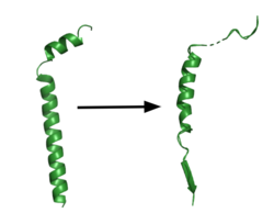
[[Image:App.png|250 px|right|thumb|'''Figure 1. APP fragment conformational change in gamma secretase.''' APP bound to gamma secretase undergoes a conformational change. The free state consists of 2 helices. The N-terminal helix unfolds into a coil and the C-terminal helix unwinds into a β-strand. This β-strand interacts with PS1 and is the site of cleavage by gamma secretase.]] | [[Image:App.png|250 px|right|thumb|'''Figure 1. APP fragment conformational change in gamma secretase.''' APP bound to gamma secretase undergoes a conformational change. The free state consists of 2 helices. The N-terminal helix unfolds into a coil and the C-terminal helix unwinds into a β-strand. This β-strand interacts with PS1 and is the site of cleavage by gamma secretase.]] | ||
| - | GS has | + | GS has been structurally characterized in the presence of both [https://en.wikipedia.org/wiki/Amyloid_precursor_protein/ APP] and Notch substrates. In each of these structures, the substrate bound in a similar location and underwent a similar structural transition upon binding to the active site of GS. Each substrate is composed of an N-terminal loop and a transmembrane helix. The peptide substrate enters the enzyme by <scene name='83/832945/App_in_gs_general/1'>lateral diffusion</scene> via the lid complex, and once in place, the TM helix of the substrate is anchored by <scene name='83/832945/Hydrophobic_interactions/2'>van der Waals contacts</scene>. Upon binding to GS, the C-terminal extracellular helix unwinds. The N-terminal end of the TM helix unwinds into a β-strand (Fig. 1). To differentiate substrates, the β-strand is often the main point of identification for the enzyme. Substrate binding induces a structural change in GS, creating two β-strands that form a β-sheet with the one β-strand of the substrate. This β-sheet is in close proximity with the active site, and guides the process of catalysis.<ref name="Zhou">PMID:30630874</ref> |
| Line 28: | Line 28: | ||
===Active Site=== | ===Active Site=== | ||
| - | The <scene name='83/832945/Asp_257_and_asp_385/12'>active site</scene> is located between TM6 and TM7 of the PS1 subunit, which is mainly hydrophilic and disordered. | + | The <scene name='83/832945/Asp_257_and_asp_385/12'>active site</scene> is located between TM6 and TM7 of the PS1 subunit, which is mainly hydrophilic and disordered. Both TM6 and TM7 contribute an aspartate residue to the active site. These two aspartates, <scene name='83/832945/Asp_257_and_asp_385/10'>Asp257 and Asp385</scene> are located approximately 10.6 A˚ apart when inactive.<ref name="Bai">PMID:26280335</ref> Substrate recognition is controlled by the closely spaced PAL sequence of <scene name='83/832945/Asp_257_and_asp_385/11'>Pro433, Ala434, and Leu435</scene>. GS becomes active upon substrate binding, when TM2 and TM6 each rotate about 15 degrees to more closely associate. Two β-strands are induced in PS1, creating an <scene name='83/832945/Beta_sheet_complex/1'>antiparallel β-sheet</scene> with the β-strand of APP.<ref name="Zhou" /> The β-strand of the substrate interacts via main chain H-bonds with the PAL sequence, stabilizing the active site. Asp257 and Asp385 hydrogen bond to each other and are located 6–7 Å away from the scissile peptide bond of the substrate, allowing catalysis to occur.<ref name="Yang" /> The cleavage site is between the helix and the N-terminal β-strand of APP. GS cleaves in 3 residue segments which is driven by the presence of three amino acid binding pockets in the active site. GS can cleave via different pathways, depending on its starting point, but the 2 most commonly used pathways produce Aβ48 and Aβ49.<ref name="Bolduc">PMID:27580372</ref>. Tripeptide cleavage starting between <scene name='83/832945/3_residues_for_cleavage/2'>Thr719 and Leu720</scene> results in Aβ48. Cleavage between <scene name='83/832945/3_residues_for_cleavage/3'>Leu720 and Val721</scene> yields Aβ49.<ref name="Zhou">PMID:30630874</ref> |
Revision as of 01:18, 21 April 2020
Gamma Secretase
| |||||||||||
References
- ↑ 1.0 1.1 Yang G, Zhou R, Shi Y. Cryo-EM structures of human gamma-secretase. Curr Opin Struct Biol. 2017 Oct;46:55-64. doi: 10.1016/j.sbi.2017.05.013. Epub, 2017 Jul 17. PMID:28628788 doi:http://dx.doi.org/10.1016/j.sbi.2017.05.013
- ↑ 2.0 2.1 2.2 2.3 Zhou R, Yang G, Guo X, Zhou Q, Lei J, Shi Y. Recognition of the amyloid precursor protein by human gamma-secretase. Science. 2019 Feb 15;363(6428). pii: science.aaw0930. doi:, 10.1126/science.aaw0930. Epub 2019 Jan 10. PMID:30630874 doi:http://dx.doi.org/10.1126/science.aaw0930
- ↑ 3.0 3.1 Bai XC, Yan C, Yang G, Lu P, Ma D, Sun L, Zhou R, Scheres SH, Shi Y. An atomic structure of human gamma-secretase. Nature. 2015 Aug 17. doi: 10.1038/nature14892. PMID:26280335 doi:http://dx.doi.org/10.1038/nature14892
- ↑ Bolduc DM, Montagna DR, Seghers MC, Wolfe MS, Selkoe DJ. The amyloid-beta forming tripeptide cleavage mechanism of gamma-secretase. Elife. 2016 Aug 31;5. doi: 10.7554/eLife.17578. PMID:27580372 doi:http://dx.doi.org/10.7554/eLife.17578
- ↑ Kumar D, Ganeshpurkar A, Kumar D, Modi G, Gupta SK, Singh SK. Secretase inhibitors for the treatment of Alzheimer's disease: Long road ahead. Eur J Med Chem. 2018 Mar 25;148:436-452. doi: 10.1016/j.ejmech.2018.02.035. Epub , 2018 Feb 15. PMID:29477076 doi:http://dx.doi.org/10.1016/j.ejmech.2018.02.035
Student Contributors
Layla Wisser
Daniel Mulawa

