We apologize for Proteopedia being slow to respond. For the past two years, a new implementation of Proteopedia has been being built. Soon, it will replace this 18-year old system. All existing content will be moved to the new system at a date that will be announced here.
User:R. Jeremy Johnson/Mitochondrial Calcium Uniporter
From Proteopedia
(Difference between revisions)
| Line 3: | Line 3: | ||
== Overview == | == Overview == | ||
| - | |||
The mitochondrial calcium uniporter (MCU) complex is the main source of entry for [https://en.wikipedia.org/wiki/Calcium calcium] ions into the [https://en.wikipedia.org/wiki/Mitochondrial_matrix mitochondrial matrix] from the [https://en.wikipedia.org/wiki/Mitochondrion#Intermembrane_space intermembrane space]. MCU channels exist in most [https://en.wikipedia.org/wiki/Eukaryote eukaryotic] life, but activity is regulated differently in each [https://en.wikipedia.org/wiki/Clade clade].<ref name="Baradaran">PMID:29995857</ref> The precise identity of the MCU wasn't discovered until 2011 and was discovered using a combination of [https://en.wikipedia.org/wiki/Nuclear_magnetic_resonance_spectroscopy NMR spectroscopy], [https://en.wikipedia.org/wiki/Transmission_electron_cryomicroscopy cryo-electron microscopy], and [https://en.wikipedia.org/wiki/X-ray_crystallography x-ray crystallography].<ref name="Woods">PMID:31869674</ref> [https://en.wikipedia.org/wiki/Cryogenic_electron_microscopy Cryoelectron microscopy] (Cryo-EM) was instrumental in understanding the complete structure of the MCU. Cryo-EM analysis provided a structural framework for understanding the mechanism by with the MCU functions.<ref name="Giorgi" /> Prior modeling of the structure was difficult because it has no apparent sequence similarity to other ion channels.<ref name="Baradaran"/> However, like other ion channels, the MCU is highly selective and efficient. The MCU has the ability to only allow calcium ions into the mitochondrial matrix at a rate of 5,000,000 ions per second even though [https://en.wikipedia.org/wiki/Potassium potassium] ions are over 100,000 times more abundant in the intermembrane space.<ref name="Baradaran"/> | The mitochondrial calcium uniporter (MCU) complex is the main source of entry for [https://en.wikipedia.org/wiki/Calcium calcium] ions into the [https://en.wikipedia.org/wiki/Mitochondrial_matrix mitochondrial matrix] from the [https://en.wikipedia.org/wiki/Mitochondrion#Intermembrane_space intermembrane space]. MCU channels exist in most [https://en.wikipedia.org/wiki/Eukaryote eukaryotic] life, but activity is regulated differently in each [https://en.wikipedia.org/wiki/Clade clade].<ref name="Baradaran">PMID:29995857</ref> The precise identity of the MCU wasn't discovered until 2011 and was discovered using a combination of [https://en.wikipedia.org/wiki/Nuclear_magnetic_resonance_spectroscopy NMR spectroscopy], [https://en.wikipedia.org/wiki/Transmission_electron_cryomicroscopy cryo-electron microscopy], and [https://en.wikipedia.org/wiki/X-ray_crystallography x-ray crystallography].<ref name="Woods">PMID:31869674</ref> [https://en.wikipedia.org/wiki/Cryogenic_electron_microscopy Cryoelectron microscopy] (Cryo-EM) was instrumental in understanding the complete structure of the MCU. Cryo-EM analysis provided a structural framework for understanding the mechanism by with the MCU functions.<ref name="Giorgi" /> Prior modeling of the structure was difficult because it has no apparent sequence similarity to other ion channels.<ref name="Baradaran"/> However, like other ion channels, the MCU is highly selective and efficient. The MCU has the ability to only allow calcium ions into the mitochondrial matrix at a rate of 5,000,000 ions per second even though [https://en.wikipedia.org/wiki/Potassium potassium] ions are over 100,000 times more abundant in the intermembrane space.<ref name="Baradaran"/> | ||
| Line 9: | Line 8: | ||
Regulation of the uptake and efflux of calcium is important to increase calcium levels enough to activate certain enzymes, but also avoid calcium overload and [https://en.wikipedia.org/wiki/Apoptosis apoptosis].<ref name="Wang"/> Mitochondrial calcium increases [http://proteopedia.org/wiki/index.php/ATP ATP] production by activating [http://proteopedia.org/wiki/index.php/Pyruvate_dehydrogenase pyruvate dehydrogenase], [https://en.wikipedia.org/wiki/Oxoglutarate_dehydrogenase_complex α-ketoglutarate dehydrogenase], and [http://proteopedia.org/wiki/index.php/Isocitrate_dehydrogenase isocitrate dehydrogenase] in the [https://en.wikipedia.org/wiki/Citric_acid_cycle Krebs cycle].<ref name="Wang"/> Therefore, deficiency of MCU leads to decrease of enzyme activity and of [https://en.wikipedia.org/wiki/Oxidative_phosphorylation oxidative phosphorylation]. | Regulation of the uptake and efflux of calcium is important to increase calcium levels enough to activate certain enzymes, but also avoid calcium overload and [https://en.wikipedia.org/wiki/Apoptosis apoptosis].<ref name="Wang"/> Mitochondrial calcium increases [http://proteopedia.org/wiki/index.php/ATP ATP] production by activating [http://proteopedia.org/wiki/index.php/Pyruvate_dehydrogenase pyruvate dehydrogenase], [https://en.wikipedia.org/wiki/Oxoglutarate_dehydrogenase_complex α-ketoglutarate dehydrogenase], and [http://proteopedia.org/wiki/index.php/Isocitrate_dehydrogenase isocitrate dehydrogenase] in the [https://en.wikipedia.org/wiki/Citric_acid_cycle Krebs cycle].<ref name="Wang"/> Therefore, deficiency of MCU leads to decrease of enzyme activity and of [https://en.wikipedia.org/wiki/Oxidative_phosphorylation oxidative phosphorylation]. | ||
| + | |||
| + | ==Structure== | ||
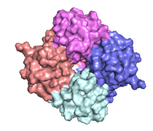
[[Image:structure.png|300 px|right|thumb|Figure 1: Structure of mitochondrial calcium uniporter colored by functional domain. The transmembrane domain is highlighted in salmon, the matrix domain in light cyan, coiled-coil domain in dark violet, and the N-terminal domain in slate blue. [https://en.wikipedia.org/wiki/Protein_Data_Bank PDB] [https://www.rcsb.org/structure/6DT0 6DT0]]] | [[Image:structure.png|300 px|right|thumb|Figure 1: Structure of mitochondrial calcium uniporter colored by functional domain. The transmembrane domain is highlighted in salmon, the matrix domain in light cyan, coiled-coil domain in dark violet, and the N-terminal domain in slate blue. [https://en.wikipedia.org/wiki/Protein_Data_Bank PDB] [https://www.rcsb.org/structure/6DT0 6DT0]]] | ||
| - | ==Structure== | ||
===Mitochondrial Calcium Uniporter Complex=== | ===Mitochondrial Calcium Uniporter Complex=== | ||
| Line 16: | Line 16: | ||
===Mitochondrial Calcium Uniporter=== | ===Mitochondrial Calcium Uniporter=== | ||
| - | |||
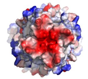
The <scene name='83/832952/Starting_scene/5'>mitochondrial calcium uniporter (MCU)</scene> is the ion channel component (Figure 1). The MCU was originally thought to be composed of [https://en.wikipedia.org/wiki/Pentamer pentamer] of five identical subunits, but it is now known to exist as a [https://en.wikipedia.org/wiki/Dimer_(chemistry) dimer] of <scene name='83/832952/Dimer_of_dimers/5'>dimers</scene> (Figure 2).<ref name="Woods">PMID:31869674</ref> The <scene name='83/837230/Ntermsymmetry/1'>dimeric</scene> organization of the MCU is described as a <scene name='83/837230/Pyramid/2'>tetrameric truncated pyramid</scene>. The protein is composed of a <scene name='83/837230/Transmembrane_domain/3'>transmembrane domain</scene>, a <scene name='83/837230/Coiled_coil/3'>coiled coil domain</scene>, and a <scene name='83/837230/Nterm/2'>N-Terminal Domain</scene> (NTD) (Figure 1).<ref name="Woods"/> The hydrophobic <scene name='83/832952/New_ones/3'>transmembrane domain</scene> is located in the inner mitochondrial membrane ([https://en.wikipedia.org/wiki/Inner_mitochondrial_membrane IMM]) and the hydrophilic coiled-coil domain exists in the mitochondrial matrix.<ref name="Baradaran"/> | The <scene name='83/832952/Starting_scene/5'>mitochondrial calcium uniporter (MCU)</scene> is the ion channel component (Figure 1). The MCU was originally thought to be composed of [https://en.wikipedia.org/wiki/Pentamer pentamer] of five identical subunits, but it is now known to exist as a [https://en.wikipedia.org/wiki/Dimer_(chemistry) dimer] of <scene name='83/832952/Dimer_of_dimers/5'>dimers</scene> (Figure 2).<ref name="Woods">PMID:31869674</ref> The <scene name='83/837230/Ntermsymmetry/1'>dimeric</scene> organization of the MCU is described as a <scene name='83/837230/Pyramid/2'>tetrameric truncated pyramid</scene>. The protein is composed of a <scene name='83/837230/Transmembrane_domain/3'>transmembrane domain</scene>, a <scene name='83/837230/Coiled_coil/3'>coiled coil domain</scene>, and a <scene name='83/837230/Nterm/2'>N-Terminal Domain</scene> (NTD) (Figure 1).<ref name="Woods"/> The hydrophobic <scene name='83/832952/New_ones/3'>transmembrane domain</scene> is located in the inner mitochondrial membrane ([https://en.wikipedia.org/wiki/Inner_mitochondrial_membrane IMM]) and the hydrophilic coiled-coil domain exists in the mitochondrial matrix.<ref name="Baradaran"/> | ||
[[Image:Nterm.png|300 px|right|thumb|Figure 2: Symmetry and organization of subunits from looking down into the uniporter from the inner mitochondrial membrane [https://en.wikipedia.org/wiki/Protein_Data_Bank PDB] [https://www.rcsb.org/structure/6DT0 6DT0]]] | [[Image:Nterm.png|300 px|right|thumb|Figure 2: Symmetry and organization of subunits from looking down into the uniporter from the inner mitochondrial membrane [https://en.wikipedia.org/wiki/Protein_Data_Bank PDB] [https://www.rcsb.org/structure/6DT0 6DT0]]] | ||
| Line 38: | Line 37: | ||
===Movement of Calcium=== | ===Movement of Calcium=== | ||
| - | |||
Cryo-EM showed three <scene name='83/832952/Starting_scene/5'>calciums</scene> in the MCU channel of roughly spherical density equally spaced 6Å apart.<ref name="Baradaran"/> Sites 1 and 2 lie within the <scene name='83/832952/Selectivity_filter/3'>selectivity filter</scene> and likely contain calcium, but site 3 could be calcium or some other small molecule.<ref name="Baradaran"/> Site 1 is positioned in the ring formed by <scene name='83/832952/Selectivity_filter_asp/2'>Asp225</scene> residues and there is a distance of 4Å between the center of the site and each [https://en.wikipedia.org/wiki/Carboxylate carboxylate group] indicating presence of water.<ref name="Baradaran"/> The second site is positioned in the ring formed by <scene name='83/832952/Selectivity_filter_glu/3'>Glu228</scene> and there is a distance of 2.8Å between the carboxylate group of each residue and the middle of the site indicating absence of water.<ref name="Baradaran"/> For transporting calcium, it is proposed that one calcium ion coordinated with water positioned in site 1 loses its water and moves to site 2 and a calcium ion moves from the IMS into site 1.<ref name="Baradaran"/> Meanwhile, a different calcium ion moves from site 2 to site 3 or the mitochondrial matrix.<ref name="Baradaran"/> | Cryo-EM showed three <scene name='83/832952/Starting_scene/5'>calciums</scene> in the MCU channel of roughly spherical density equally spaced 6Å apart.<ref name="Baradaran"/> Sites 1 and 2 lie within the <scene name='83/832952/Selectivity_filter/3'>selectivity filter</scene> and likely contain calcium, but site 3 could be calcium or some other small molecule.<ref name="Baradaran"/> Site 1 is positioned in the ring formed by <scene name='83/832952/Selectivity_filter_asp/2'>Asp225</scene> residues and there is a distance of 4Å between the center of the site and each [https://en.wikipedia.org/wiki/Carboxylate carboxylate group] indicating presence of water.<ref name="Baradaran"/> The second site is positioned in the ring formed by <scene name='83/832952/Selectivity_filter_glu/3'>Glu228</scene> and there is a distance of 2.8Å between the carboxylate group of each residue and the middle of the site indicating absence of water.<ref name="Baradaran"/> For transporting calcium, it is proposed that one calcium ion coordinated with water positioned in site 1 loses its water and moves to site 2 and a calcium ion moves from the IMS into site 1.<ref name="Baradaran"/> Meanwhile, a different calcium ion moves from site 2 to site 3 or the mitochondrial matrix.<ref name="Baradaran"/> | ||
===Mutations=== | ===Mutations=== | ||
| - | |||
A number of mutations completely eliminate calcium uptake by the MCU. For example, mutation of [https://en.wikipedia.org/wiki/Tryptophan Trp224], [https://en.wikipedia.org/wiki/Aspartic_acid Asp225], [https://en.wikipedia.org/wiki/Glutamic_acid Glu228], or [https://en.wikipedia.org/wiki/Proline Pro229] of the WDXXEP motif altered the highly conserved selectivity filter and completely eliminated calcium uptake.<ref name="Baradaran"/><ref name="Fan"/> Even mutating Glu228 to an aspartate significantly changed the dimensions of the pore and inhibited uptake of calcium.<ref name="Baradaran"/> However, mutation of either X residue was not detrimental to calcium uptake.<ref name="Baradaran"/> Furthermore, mutation of the tyrosine residue directly below the selectivity filter substantially impaired calcium intake and proper protein folding.<ref name="Fan"/> The residue on TM1 that affected calcium uptake the most in human MCU was Trp317 (analogous to <scene name='83/832952/New_ones/7'>Trp210</scene> in ''C. europaea'') which has a side chain constituting a primary contact point between TM1 and TM2.<ref name="Fan"/> Mutation of human MCU Phe326 (analogous to <scene name='83/832952/New_ones/8'>Phe218</scene> in ''C. europaea'') or Gly331 of the TM1-TM2 linker (<scene name='83/832952/New_ones/9'>Gly223</scene> in ''C. europaea'') affected the linker conformation and configuration of the pore entrance and impaired calcium intake.<ref name="Fan"/> | A number of mutations completely eliminate calcium uptake by the MCU. For example, mutation of [https://en.wikipedia.org/wiki/Tryptophan Trp224], [https://en.wikipedia.org/wiki/Aspartic_acid Asp225], [https://en.wikipedia.org/wiki/Glutamic_acid Glu228], or [https://en.wikipedia.org/wiki/Proline Pro229] of the WDXXEP motif altered the highly conserved selectivity filter and completely eliminated calcium uptake.<ref name="Baradaran"/><ref name="Fan"/> Even mutating Glu228 to an aspartate significantly changed the dimensions of the pore and inhibited uptake of calcium.<ref name="Baradaran"/> However, mutation of either X residue was not detrimental to calcium uptake.<ref name="Baradaran"/> Furthermore, mutation of the tyrosine residue directly below the selectivity filter substantially impaired calcium intake and proper protein folding.<ref name="Fan"/> The residue on TM1 that affected calcium uptake the most in human MCU was Trp317 (analogous to <scene name='83/832952/New_ones/7'>Trp210</scene> in ''C. europaea'') which has a side chain constituting a primary contact point between TM1 and TM2.<ref name="Fan"/> Mutation of human MCU Phe326 (analogous to <scene name='83/832952/New_ones/8'>Phe218</scene> in ''C. europaea'') or Gly331 of the TM1-TM2 linker (<scene name='83/832952/New_ones/9'>Gly223</scene> in ''C. europaea'') affected the linker conformation and configuration of the pore entrance and impaired calcium intake.<ref name="Fan"/> | ||
==Regulation and Inhibition== | ==Regulation and Inhibition== | ||
| - | + | [[Image:Ruthenium_Inhibitors.jpg|220 px|right|thumb|Figure 4: Structures of the ruthenium-based inhibitors of the MCU. Created using ChemDraw Professional 16.0]] | |
| - | [[Image:Ruthenium_Inhibitors.jpg| | + | |
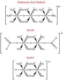
The most well-known and commonly used inhibitor of the MCU is [https://en.wikipedia.org/wiki/Ruthenium_red ruthenium red] (RuRed).<ref name="Woods"/> RuRed is a trinuclear, oxo-bridged complex that effectively inhibits calcium uptake without affecting mitochondrial respiration or calcium efflux.<ref name="Woods"/> The disadvantage of ruthenium red is its challenging purification.<ref name="Woods"/> Interestingly, an impure version of RuRed, termed [https://en.wikipedia.org/wiki/Ru360 Ru360], was found to be the active component of RuRed and thus another good inhibitor of the MCU.<ref name="Woods"/> Ru360 is a binuclear, oxo-bridged complex with a similar structure to that of RuRed.<ref name="Woods"/> The only flaw with Ru360 was that it showed low cell permeability, so Ru265 was developed and had twice the cell permeability of Ru360.<ref name="Woods"/> Ru265 possesses two bridged Ru centers bridged by a nitride ligand.<ref name="Woods"/> | The most well-known and commonly used inhibitor of the MCU is [https://en.wikipedia.org/wiki/Ruthenium_red ruthenium red] (RuRed).<ref name="Woods"/> RuRed is a trinuclear, oxo-bridged complex that effectively inhibits calcium uptake without affecting mitochondrial respiration or calcium efflux.<ref name="Woods"/> The disadvantage of ruthenium red is its challenging purification.<ref name="Woods"/> Interestingly, an impure version of RuRed, termed [https://en.wikipedia.org/wiki/Ru360 Ru360], was found to be the active component of RuRed and thus another good inhibitor of the MCU.<ref name="Woods"/> Ru360 is a binuclear, oxo-bridged complex with a similar structure to that of RuRed.<ref name="Woods"/> The only flaw with Ru360 was that it showed low cell permeability, so Ru265 was developed and had twice the cell permeability of Ru360.<ref name="Woods"/> Ru265 possesses two bridged Ru centers bridged by a nitride ligand.<ref name="Woods"/> | ||
| Line 53: | Line 49: | ||
==Medical Relevance== | ==Medical Relevance== | ||
| - | |||
The MCU has a large role in disease because of its effect on apoptosis and cell signaling. The overload of the mitochondrial matrix with calcium leads to release of [https://en.wikipedia.org/wiki/Cytochrome_c cytochrome c], overproduction of [https://en.wikipedia.org/wiki/Reactive_oxygen_species reactive oxygen species], mitochondrial swelling, and the opening of the mitochondrial permeability transition pore ([https://en.wikipedia.org/wiki/Mitochondrial_permeability_transition_pore mPTP]) which all lead to apoptotic cell death.<ref name="Woods"/> This connection between mitochondrial calcium and apoptosis makes the MCU dysregulation a large contributor to cell death and disease. Calcium machinery in the mitochondria are targets for [https://en.wikipedia.org/wiki/Oncogene proto-oncogenes] and [https://en.wikipedia.org/wiki/Tumor_suppressor tumor suppressors] for this very reason.<ref name="Giorgi"/> Apoptosis can either be induced or repressed. Furthermore, external stimuli can activate receptors in the endoplasmic reticulum that release calcium and activate signal transductions.<ref name="Wang"/> Sequestration of calcium in the mitochondria is vital to shut down these activations, so any impact in movement of calcium ions can cause a wide variety of diseases.<ref name="Wang"/> | The MCU has a large role in disease because of its effect on apoptosis and cell signaling. The overload of the mitochondrial matrix with calcium leads to release of [https://en.wikipedia.org/wiki/Cytochrome_c cytochrome c], overproduction of [https://en.wikipedia.org/wiki/Reactive_oxygen_species reactive oxygen species], mitochondrial swelling, and the opening of the mitochondrial permeability transition pore ([https://en.wikipedia.org/wiki/Mitochondrial_permeability_transition_pore mPTP]) which all lead to apoptotic cell death.<ref name="Woods"/> This connection between mitochondrial calcium and apoptosis makes the MCU dysregulation a large contributor to cell death and disease. Calcium machinery in the mitochondria are targets for [https://en.wikipedia.org/wiki/Oncogene proto-oncogenes] and [https://en.wikipedia.org/wiki/Tumor_suppressor tumor suppressors] for this very reason.<ref name="Giorgi"/> Apoptosis can either be induced or repressed. Furthermore, external stimuli can activate receptors in the endoplasmic reticulum that release calcium and activate signal transductions.<ref name="Wang"/> Sequestration of calcium in the mitochondria is vital to shut down these activations, so any impact in movement of calcium ions can cause a wide variety of diseases.<ref name="Wang"/> | ||
===Neurodegenerative Disorders=== | ===Neurodegenerative Disorders=== | ||
| - | |||
Disruption in calcium homeostasis leads to a wide range of [https://en.wikipedia.org/wiki/Neurodegeneration neurodegenerative disorders]. The MCU complex has been identified to play a large role in [https://en.wikipedia.org/wiki/Neuromuscular_disease neuromuscular disease] because of a loss or mutation of MICU1.<ref name="Woods"/> This causes [https://en.wikipedia.org/wiki/Myopathy myopathy], learning difficulties, and progressive movement disorders which can be lethal.<ref name="Woods"/> In [https://en.wikipedia.org/wiki/Alzheimer%27s_disease Alzheimer's disease], the buildup of [https://en.wikipedia.org/wiki/Amyloid_beta amyloid-β] plaques in the brain leads to increased calcium uptake in [https://en.wikipedia.org/wiki/Neuron neurons] and cell death.<ref name="Woods"/> Similarly, in early onset [https://en.wikipedia.org/wiki/Parkinson%27s_disease Parkinson's disease], degradation of MICU1 by the protein ligase [http://proteopedia.org/wiki/index.php/Parkin Parkin] leads to increased mitochondrial calcium uptake, overload, and death.<ref name="Woods"/> Finally, disrupted glutamate homeostasis in [https://en.wikipedia.org/wiki/Astrocyte astrocytes] and neurons leads to calcium overload and cell death via [https://en.wikipedia.org/wiki/Excitotoxicity excitotoxicity] in Amyotrophic Lateral Sclerosis ([https://en.wikipedia.org/wiki/Amyotrophic_lateral_sclerosis ALS]).<ref name="Woods"/> | Disruption in calcium homeostasis leads to a wide range of [https://en.wikipedia.org/wiki/Neurodegeneration neurodegenerative disorders]. The MCU complex has been identified to play a large role in [https://en.wikipedia.org/wiki/Neuromuscular_disease neuromuscular disease] because of a loss or mutation of MICU1.<ref name="Woods"/> This causes [https://en.wikipedia.org/wiki/Myopathy myopathy], learning difficulties, and progressive movement disorders which can be lethal.<ref name="Woods"/> In [https://en.wikipedia.org/wiki/Alzheimer%27s_disease Alzheimer's disease], the buildup of [https://en.wikipedia.org/wiki/Amyloid_beta amyloid-β] plaques in the brain leads to increased calcium uptake in [https://en.wikipedia.org/wiki/Neuron neurons] and cell death.<ref name="Woods"/> Similarly, in early onset [https://en.wikipedia.org/wiki/Parkinson%27s_disease Parkinson's disease], degradation of MICU1 by the protein ligase [http://proteopedia.org/wiki/index.php/Parkin Parkin] leads to increased mitochondrial calcium uptake, overload, and death.<ref name="Woods"/> Finally, disrupted glutamate homeostasis in [https://en.wikipedia.org/wiki/Astrocyte astrocytes] and neurons leads to calcium overload and cell death via [https://en.wikipedia.org/wiki/Excitotoxicity excitotoxicity] in Amyotrophic Lateral Sclerosis ([https://en.wikipedia.org/wiki/Amyotrophic_lateral_sclerosis ALS]).<ref name="Woods"/> | ||
===Diabetes=== | ===Diabetes=== | ||
| - | |||
Calcium homeostasis misregulation has also been proven to be instrumental in [https://en.wikipedia.org/wiki/Obesity obesity], [https://en.wikipedia.org/wiki/Insulin_resistance insulin resistance], and [https://en.wikipedia.org/wiki/Type_2_diabetes type-II diabetes].<ref name="Wang"/> The intracellular calcium concentrations in primary [https://en.wikipedia.org/wiki/Adipocyte adipocytes] from obese human subjects has been found to be elevated.<ref name="Wang"/> Any inhibition of downstream calcium signaling could decrease movement of the [http://proteopedia.org/wiki/index.php/GLUT4 GLUT4] glucose transporter and glucose uptake.<ref name="Wang"/> Additionally, [https://en.wikipedia.org/wiki/Ablation ablation] of MCU in [https://en.wikipedia.org/wiki/Beta_cell β-cells] in the [https://en.wikipedia.org/wiki/Pancreas pancreas] demonstrated a decrease in cellular ATP concentration following glucose stimulation which resulted in decreased glucose-stimulated insulin secretion.<ref name="Wang"/> Furthermore, MAMs have been shown to cause glucose intolerance and mitochondrial dysfunction in primary [https://en.wikipedia.org/wiki/Hepatocyte hepatocytes] in mice.<ref name="Wang"/> Subsequent reinforcement of these MAMs has been shown to increase insulin sensitivity and glucose homeostasis.<ref name="Wang"/> | Calcium homeostasis misregulation has also been proven to be instrumental in [https://en.wikipedia.org/wiki/Obesity obesity], [https://en.wikipedia.org/wiki/Insulin_resistance insulin resistance], and [https://en.wikipedia.org/wiki/Type_2_diabetes type-II diabetes].<ref name="Wang"/> The intracellular calcium concentrations in primary [https://en.wikipedia.org/wiki/Adipocyte adipocytes] from obese human subjects has been found to be elevated.<ref name="Wang"/> Any inhibition of downstream calcium signaling could decrease movement of the [http://proteopedia.org/wiki/index.php/GLUT4 GLUT4] glucose transporter and glucose uptake.<ref name="Wang"/> Additionally, [https://en.wikipedia.org/wiki/Ablation ablation] of MCU in [https://en.wikipedia.org/wiki/Beta_cell β-cells] in the [https://en.wikipedia.org/wiki/Pancreas pancreas] demonstrated a decrease in cellular ATP concentration following glucose stimulation which resulted in decreased glucose-stimulated insulin secretion.<ref name="Wang"/> Furthermore, MAMs have been shown to cause glucose intolerance and mitochondrial dysfunction in primary [https://en.wikipedia.org/wiki/Hepatocyte hepatocytes] in mice.<ref name="Wang"/> Subsequent reinforcement of these MAMs has been shown to increase insulin sensitivity and glucose homeostasis.<ref name="Wang"/> | ||
===Heart Failure=== | ===Heart Failure=== | ||
| - | |||
Calcium overload in the mitochondria of cardiac cells lead to [https://en.wikipedia.org/wiki/Apoptosis apoptotic] cardiac cell death. Calcium governs [https://en.wikipedia.org/wiki/Cardiac_excitation-contraction_coupling excitation contraction coupling] (EC coupling) of the cardiac muscles, which creates the ATP needed to power the contraction during heart beats. The increase in mitochondrial Ca2+ concentration is essential for the functioning of this muscle contraction. Mitochondrial Ca2+ overload, though, leads to necrotic cardiac cell death and can be targeted with regulation of the MCU. An example of treatment would be with the use of Ru360 to inhibit the uptake of Ca2+ ions into the mitochondria. <ref name="Giorgi" /> | Calcium overload in the mitochondria of cardiac cells lead to [https://en.wikipedia.org/wiki/Apoptosis apoptotic] cardiac cell death. Calcium governs [https://en.wikipedia.org/wiki/Cardiac_excitation-contraction_coupling excitation contraction coupling] (EC coupling) of the cardiac muscles, which creates the ATP needed to power the contraction during heart beats. The increase in mitochondrial Ca2+ concentration is essential for the functioning of this muscle contraction. Mitochondrial Ca2+ overload, though, leads to necrotic cardiac cell death and can be targeted with regulation of the MCU. An example of treatment would be with the use of Ru360 to inhibit the uptake of Ca2+ ions into the mitochondria. <ref name="Giorgi" /> | ||
</StructureSection> | </StructureSection> | ||
Revision as of 19:42, 8 May 2020
Mitochondrial Calcium Uniporter (MCU) Complex
| |||||||||||
References
- ↑ 1.00 1.01 1.02 1.03 1.04 1.05 1.06 1.07 1.08 1.09 1.10 1.11 1.12 1.13 1.14 1.15 1.16 1.17 1.18 1.19 1.20 1.21 1.22 1.23 1.24 1.25 1.26 1.27 Baradaran R, Wang C, Siliciano AF, Long SB. Cryo-EM structures of fungal and metazoan mitochondrial calcium uniporters. Nature. 2018 Jul 11. pii: 10.1038/s41586-018-0331-8. doi:, 10.1038/s41586-018-0331-8. PMID:29995857 doi:http://dx.doi.org/10.1038/s41586-018-0331-8
- ↑ 2.00 2.01 2.02 2.03 2.04 2.05 2.06 2.07 2.08 2.09 2.10 2.11 2.12 2.13 2.14 2.15 2.16 2.17 2.18 Woods JJ, Wilson JJ. Inhibitors of the mitochondrial calcium uniporter for the treatment of disease. Curr Opin Chem Biol. 2019 Dec 20;55:9-18. doi: 10.1016/j.cbpa.2019.11.006. PMID:31869674 doi:http://dx.doi.org/10.1016/j.cbpa.2019.11.006
- ↑ 3.0 3.1 3.2 3.3 3.4 3.5 3.6 3.7 3.8 Giorgi C, Marchi S, Pinton P. The machineries, regulation and cellular functions of mitochondrial calcium. Nat Rev Mol Cell Biol. 2018 Nov;19(11):713-730. doi: 10.1038/s41580-018-0052-8. PMID:30143745 doi:http://dx.doi.org/10.1038/s41580-018-0052-8
- ↑ 4.00 4.01 4.02 4.03 4.04 4.05 4.06 4.07 4.08 4.09 4.10 4.11 4.12 4.13 4.14 4.15 4.16 4.17 Wang CH, Wei YH. Role of mitochondrial dysfunction and dysregulation of Ca(2+) homeostasis in the pathophysiology of insulin resistance and type 2 diabetes. J Biomed Sci. 2017 Sep 7;24(1):70. doi: 10.1186/s12929-017-0375-3. PMID:28882140 doi:http://dx.doi.org/10.1186/s12929-017-0375-3
- ↑ 5.00 5.01 5.02 5.03 5.04 5.05 5.06 5.07 5.08 5.09 5.10 5.11 5.12 Fan C, Fan M, Orlando BJ, Fastman NM, Zhang J, Xu Y, Chambers MG, Xu X, Perry K, Liao M, Feng L. X-ray and cryo-EM structures of the mitochondrial calcium uniporter. Nature. 2018 Jul 11. pii: 10.1038/s41586-018-0330-9. doi:, 10.1038/s41586-018-0330-9. PMID:29995856 doi:http://dx.doi.org/10.1038/s41586-018-0330-9
- ↑ Yoo J, Wu M, Yin Y, Herzik MA Jr, Lander GC, Lee SY. Cryo-EM structure of a mitochondrial calcium uniporter. Science. 2018 Jun 28. pii: science.aar4056. doi: 10.1126/science.aar4056. PMID:29954988 doi:http://dx.doi.org/10.1126/science.aar4056
Student Contributors
Ryan Heumann
Lizzy Ratz
Holly Rowe
Madi Summers
Rieser Wells


