We apologize for Proteopedia being slow to respond. For the past two years, a new implementation of Proteopedia has been being built. Soon, it will replace this 18-year old system. All existing content will be moved to the new system at a date that will be announced here.
1dxx
From Proteopedia
(Difference between revisions)
| Line 1: | Line 1: | ||
| - | [[Image:1dxx. | + | {{Seed}} |
| + | [[Image:1dxx.png|left|200px]] | ||
<!-- | <!-- | ||
| Line 9: | Line 10: | ||
{{STRUCTURE_1dxx| PDB=1dxx | SCENE= }} | {{STRUCTURE_1dxx| PDB=1dxx | SCENE= }} | ||
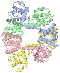
| - | + | ===N-TERMINAL ACTIN-BINDING DOMAIN OF HUMAN DYSTROPHIN=== | |
| - | + | <!-- | |
| - | + | The line below this paragraph, {{ABSTRACT_PUBMED_10801490}}, adds the Publication Abstract to the page | |
| + | (as it appears on PubMed at http://www.pubmed.gov), where 10801490 is the PubMed ID number. | ||
| + | --> | ||
| + | {{ABSTRACT_PUBMED_10801490}} | ||
==Disease== | ==Disease== | ||
| Line 34: | Line 38: | ||
[[Category: Muscular dystrophy]] | [[Category: Muscular dystrophy]] | ||
[[Category: Utrophin]] | [[Category: Utrophin]] | ||
| - | ''Page seeded by [http://oca.weizmann.ac.il/oca OCA ] on | + | |
| + | ''Page seeded by [http://oca.weizmann.ac.il/oca OCA ] on Mon Jun 30 23:47:12 2008'' | ||
Revision as of 20:47, 30 June 2008
Contents |
N-TERMINAL ACTIN-BINDING DOMAIN OF HUMAN DYSTROPHIN
Template:ABSTRACT PUBMED 10801490
Disease
Known disease associated with this structure: Becker muscular dystrophy OMIM:[300377], Cardiomyopathy, dilated, 3B OMIM:[300377], Duchenne muscular dystrophy OMIM:[300377]
About this Structure
1DXX is a Single protein structure of sequence from Homo sapiens. Full crystallographic information is available from OCA.
Reference
The structure of the N-terminal actin-binding domain of human dystrophin and how mutations in this domain may cause Duchenne or Becker muscular dystrophy., Norwood FL, Sutherland-Smith AJ, Keep NH, Kendrick-Jones J, Structure. 2000 May 15;8(5):481-91. PMID:10801490
Page seeded by OCA on Mon Jun 30 23:47:12 2008

