This old version of Proteopedia is provided for student assignments while the new version is undergoing repairs. Content and edits done in this old version of Proteopedia after March 1, 2026 will eventually be lost when it is retired in about June of 2026.
Apply for new accounts at the new Proteopedia. Your logins will work in both the old and new versions.
User:Leanne Price/Sandbox 1
From Proteopedia
| Line 18: | Line 18: | ||
==Structure== | ==Structure== | ||
| - | DGAT consists of two domains, one cytoplasmic and one luminal. The cytoplasmic domain interacts with the interior of the cell and relays signals. The luminal domain senses misfolded proteins. The structure of DGAT consists of two protein chains, one ligand, two polymers, eighteen <scene name='87/877601/Transmembrane_helices/2'>alpha helices</scene> and zero beta sheets. The transmembrane helices form a large central cavity within the membrane that opens to the bilayer via a wide lateral gate. Through openings on the cytosolic and luminal sides of DGAT, this central cavity is also accessible. The majority of the transmembrane helices present within the structure also form a concave-shaped ridge on either side of the membrane. These aspects of the domain structure are deemed as the 'MBOAT core'. Within this core, a tunnel-like region, similar to a binding pocket, is also present. Access to the active site of DGAT by substrates is done through the lateral gate, which lies on the ER lumen side, within the membrane. This tunnel-like region is referred to as the <scene name='87/877628/Cytosolic_s_enteranceide/1'>cytosolic, or C, tunnel</scene>. | + | DGAT consists of two domains, one cytoplasmic and one luminal. The cytoplasmic domain interacts with the interior of the cell and relays signals. The luminal domain senses misfolded proteins. The structure of DGAT consists of two protein chains, one ligand, two polymers, eighteen <scene name='87/877601/Transmembrane_helices/2'>alpha helices</scene> and zero beta sheets. The transmembrane helices form a large central cavity within the membrane that opens to the bilayer via a wide lateral gate. Through openings on the cytosolic and luminal sides of DGAT, this central cavity is also accessible. The majority of the transmembrane helices present within the structure also form a concave-shaped ridge on either side of the membrane. These aspects of the domain structure are deemed as the 'MBOAT core'. Within this core, a tunnel-like region, similar to a binding pocket, is also present. Access to the active site of DGAT by substrates is done through the lateral gate, which lies on the ER lumen side, within the membrane. This tunnel-like region is referred to as the <scene name='87/877628/Cytosolic_s_enteranceide/1'>cytosolic, or C, tunnel</scene>.<ref name="Sui" /ref>. |
[[Image:C_tunnel.PNG|400 px|left|thumb|Figure 3. Shows the location of the lateral gate as the entrance to the cytosolic tunnel via the ER lumen side of the membrane. ]] | [[Image:C_tunnel.PNG|400 px|left|thumb|Figure 3. Shows the location of the lateral gate as the entrance to the cytosolic tunnel via the ER lumen side of the membrane. ]] | ||
| Line 28: | Line 28: | ||
===Tunnels=== | ===Tunnels=== | ||
| - | DGAT consists of 3 tunnels, a cytosolic tunnel, an ER-luminal funnel, and a membrane-embedded lateral gate. The cytosolic tunnel is the site of acyl-CoA binding, with the CoA group pointing at the cytosolic face and its acyl chain pointing towards the endoplasmic reticulum lumen. DAG then enters via the lateral gate on the luminal side via the lateral gate where it can then access the active site. The resulting product can then be released to either side of the membrane. | + | DGAT consists of 3 tunnels, a cytosolic tunnel, an ER-luminal funnel, and a membrane-embedded lateral gate. The cytosolic tunnel is the site of acyl-CoA binding, with the CoA group pointing at the cytosolic face and its acyl chain pointing towards the endoplasmic reticulum lumen. DAG then enters via the lateral gate on the luminal side via the lateral gate where it can then access the active site. The resulting product can then be released to either side of the membrane. <ref name="Sui">PMID:32433611</ref> |
===Active Site=== | ===Active Site=== | ||
| Line 63: | Line 63: | ||
<ref name="Human Protein Atlas">https://www.proteinatlas.org/ENSG00000185000-DGAT1/pathology</ref> | <ref name="Human Protein Atlas">https://www.proteinatlas.org/ENSG00000185000-DGAT1/pathology</ref> | ||
| - | + | ||
<references/> | <references/> | ||
Revision as of 19:12, 13 April 2021
Diacylglycerol Acyltransferase
| |||||||||||


References
- ↑ 1.0 1.1 .
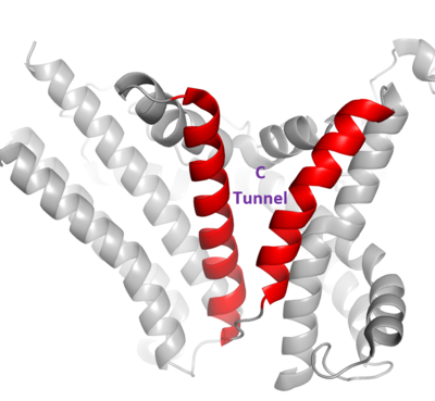 Figure 3. Shows the location of the lateral gate as the entrance to the cytosolic tunnel via the ER lumen side of the membrane.
The DGAT dimer structure is formed primarily through many hydrogen-bonding interactions between the first 20 resolved residues (His69-Gly87). Hydrophobic interactions of the transmembrane helix region (Phe82-Ile98) with the other monomer also support the dimer structure formation. Additionally, there are four phospholipids present at the dimer interface that have been thought to contribute to the interactions between DGAT monomers.
Tunnels
DGAT consists of 3 tunnels, a cytosolic tunnel, an ER-luminal funnel, and a membrane-embedded lateral gate. The cytosolic tunnel is the site of acyl-CoA binding, with the CoA group pointing at the cytosolic face and its acyl chain pointing towards the endoplasmic reticulum lumen. DAG then enters via the lateral gate on the luminal side via the lateral gate where it can then access the active site. The resulting product can then be released to either side of the membrane. <ref>PMID:32433611</li> <li id="cite_note-Human_Protein_Atlas-1">↑ <sup>[[#cite_ref-Human_Protein_Atlas_1-0|2.0]]</sup> <sup>[[#cite_ref-Human_Protein_Atlas_1-1|2.1]]</sup> https://www.proteinatlas.org/ENSG00000185000-DGAT1/pathology</li>
<li id="cite_note-Wang-2">[[#cite_ref-Wang_2-0|↑]] Wang L, Qian H, Nian Y, Han Y, Ren Z, Zhang H, Hu L, Prasad BVV, Laganowsky A, Yan N, Zhou M. Structure and mechanism of human diacylglycerol O-acyltransferase 1. Nature. 2020 May;581(7808):329-332. doi: 10.1038/s41586-020-2280-2. Epub 2020 May, 13. PMID:[http://www.ncbi.nlm.nih.gov/pubmed/32433610 32433610] doi:[http://dx.doi.org/10.1038/s41586-020-2280-2 http://dx.doi.org/10.1038/s41586-020-2280-2]</li></ol></ref>

Student Contributors
- Justin Smith
- Eloi Bigirimana
- Leanne Price
