User:Hannah Wright/Sandbox 1
From Proteopedia
(Difference between revisions)
| Line 35: | Line 35: | ||
====Mechanism==== | ====Mechanism==== | ||
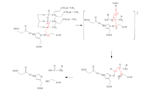
[[Image:MechFinalAllison.png|700 px|]] | [[Image:MechFinalAllison.png|700 px|]] | ||
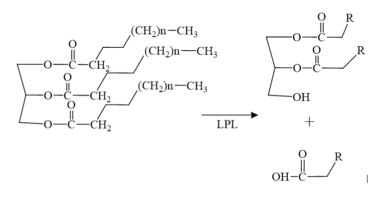
| - | The triglyceride binds to LPL’s lipid-binding region in an open lid conformation. | + | 1. The triglyceride binds to LPL’s lipid-binding region in an open lid conformation. |
| - | The oxygen on S159 is made more nucleophilic. This happens via histidine hydrogen bonding with the hydrogen on S159’s alcohol group. | + | 2. The oxygen on S159 is made more nucleophilic. This happens via histidine hydrogen bonding with the hydrogen on S159’s alcohol group. |
| - | The nucleophilic oxygen attacks the carbonyl carbon of one of the fatty acid chains. | + | 3. The nucleophilic oxygen attacks the carbonyl carbon of one of the fatty acid chains. |
| - | This pushes electrons up onto the carbonyl oxygen, creating a tetrahedral intermediate. This is the oxyanion hole which is stabilized by main chain nitrogen atoms of W82 and L160. | + | 4. This pushes electrons up onto the carbonyl oxygen, creating a tetrahedral intermediate. This is the oxyanion hole which is stabilized by main chain nitrogen atoms of W82 and L160. |
| - | One of the lone pairs of the oxygen (in the oxyanion hole) creates a double bond carbon. | + | 5. One of the lone pairs of the oxygen (in the oxyanion hole) creates a double bond carbon. |
| - | The oxygen-carbon bond between the single fatty acid chain and the diglyceride is cleaved. | + | 6. The oxygen-carbon bond between the single fatty acid chain and the diglyceride is cleaved. |
| - | H268 hydrogen bonds water, making the oxygen a better nucleophile. Water attacks the carbonyl carbon. | + | 7. H268 hydrogen bonds water, making the oxygen a better nucleophile. Water attacks the carbonyl carbon. |
| - | The carboxylic acid is formed and the S159 bond is cleaved and re-protonated via H268. | + | 8. The carboxylic acid is formed and the S159 bond is cleaved and re-protonated via H268. |
| - | The active site is now back in its original state. | + | 9. The active site is now back in its original state. |
===Inhibitors=== | ===Inhibitors=== | ||
This <scene name='87/878236/Active_site_w_inhibitor_bound/4'>inhibitor</scene>, M3D shown in a peach color, was a vital piece in unraveling the correct structure of LPL. With the inhibitor bound, LPL’s active site and lid region become visible and crystallizable, thus this inhibitor is what allowed the first complete crystal structure of LPL to come into existence. This inhibitor also revealed the correct orientation of H268, a residue involved in the catalytic triad. Originally, the hydrogen bonding did not align with substrates in the original crystal structure; however, when this inhibitor was bound, the H268 was flipped, thus aligning the hydrogen bonds in the correct orientation. | This <scene name='87/878236/Active_site_w_inhibitor_bound/4'>inhibitor</scene>, M3D shown in a peach color, was a vital piece in unraveling the correct structure of LPL. With the inhibitor bound, LPL’s active site and lid region become visible and crystallizable, thus this inhibitor is what allowed the first complete crystal structure of LPL to come into existence. This inhibitor also revealed the correct orientation of H268, a residue involved in the catalytic triad. Originally, the hydrogen bonding did not align with substrates in the original crystal structure; however, when this inhibitor was bound, the H268 was flipped, thus aligning the hydrogen bonds in the correct orientation. | ||
| Line 50: | Line 50: | ||
===Mutations=== | ===Mutations=== | ||
====D201V==== | ====D201V==== | ||
| - | <scene name='87/877636/D201_mutation/ | + | <scene name='87/877636/D201_mutation/10'>D201V</scene> is a mutation that is found to cause chylomicronemia. Chylomicronemia is when the body cannot break down lipids properly. This leads to their build-up in the body causing high levels of triglycerides in the body. The carboxyl side chain of aspartate 201 is one of the coordination sites for the calcium ion of LPL. The mutation to hydrophobic valine means the loss of this coordination site (reference Birrane). This mutation adversely affects the folding of LPL and thus affects the secretion of LPL, overall decreasing the activity of LPL (reference Birrane). |
| + | |||
====M404R==== | ====M404R==== | ||
| - | <scene name='87/877636/M404r_1/ | + | <scene name='87/877636/M404r_1/2'>M404R</scene> is a mutation found within LPL that caused chylomicronemia in patients. The hydrophobic methionine is mutated to the larger and charged side chain of arginine. Originally it was thought to impact LPL secretion from cells. It was found that the M404R does not affect LPL secretion (reference Birrane). M404R interacts with the hydrophobic pocket of GPIHBP1’s finger 3 of its 3 fingered domain (V121, E122, T124, V126). The large, charged arginine repelled the hydrophobic pocket and does not fit well. This prevents proper binding and formation of the LPL-GPIHBP1 complex (reference Birrane). |
== Relevance == | == Relevance == | ||
===Hypertriglycerademia=== | ===Hypertriglycerademia=== | ||
Revision as of 18:56, 20 April 2021
Lipoprotein Lipase (LPL) complexed with GPIHBP1
| |||||||||||
References
- ↑ Birrane G, Beigneux AP, Dwyer B, Strack-Logue B, Kristensen KK, Francone OL, Fong LG, Mertens HDT, Pan CQ, Ploug M, Young SG, Meiyappan M. Structure of the lipoprotein lipase-GPIHBP1 complex that mediates plasma triglyceride hydrolysis. Proc Natl Acad Sci U S A. 2018 Dec 17. pii: 1817984116. doi:, 10.1073/pnas.1817984116. PMID:30559189 doi:http://dx.doi.org/10.1073/pnas.1817984116
Student/Contributors
- Ashrey Burely
- Allison Welz
- Hannah Wright


