1gm5
From Proteopedia
(Difference between revisions)
| Line 3: | Line 3: | ||
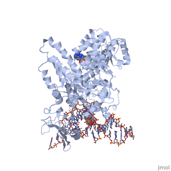
<StructureSection load='1gm5' size='340' side='right'caption='[[1gm5]], [[Resolution|resolution]] 3.24Å' scene=''> | <StructureSection load='1gm5' size='340' side='right'caption='[[1gm5]], [[Resolution|resolution]] 3.24Å' scene=''> | ||
== Structural highlights == | == Structural highlights == | ||
| - | <table><tr><td colspan='2'>[[1gm5]] is a 4 chain structure with sequence from [https://en.wikipedia.org/wiki/ | + | <table><tr><td colspan='2'>[[1gm5]] is a 4 chain structure with sequence from [https://en.wikipedia.org/wiki/Thermotoga_maritima Thermotoga maritima]. Full crystallographic information is available from [http://oca.weizmann.ac.il/oca-bin/ocashort?id=1GM5 OCA]. For a <b>guided tour on the structure components</b> use [https://proteopedia.org/fgij/fg.htm?mol=1GM5 FirstGlance]. <br> |
| - | </td></tr><tr id='ligand'><td class="sblockLbl"><b>[[Ligand|Ligands:]]</b></td><td class="sblockDat" id="ligandDat"><scene name='pdbligand=ADP:ADENOSINE-5-DIPHOSPHATE'>ADP</scene>, <scene name='pdbligand=MG:MAGNESIUM+ION'>MG</scene></td></tr> | + | </td></tr><tr id='method'><td class="sblockLbl"><b>[[Empirical_models|Method:]]</b></td><td class="sblockDat" id="methodDat">X-ray diffraction, [[Resolution|Resolution]] 3.24Å</td></tr> |
| + | <tr id='ligand'><td class="sblockLbl"><b>[[Ligand|Ligands:]]</b></td><td class="sblockDat" id="ligandDat"><scene name='pdbligand=ADP:ADENOSINE-5-DIPHOSPHATE'>ADP</scene>, <scene name='pdbligand=MG:MAGNESIUM+ION'>MG</scene></td></tr> | ||
<tr id='resources'><td class="sblockLbl"><b>Resources:</b></td><td class="sblockDat"><span class='plainlinks'>[https://proteopedia.org/fgij/fg.htm?mol=1gm5 FirstGlance], [http://oca.weizmann.ac.il/oca-bin/ocaids?id=1gm5 OCA], [https://pdbe.org/1gm5 PDBe], [https://www.rcsb.org/pdb/explore.do?structureId=1gm5 RCSB], [https://www.ebi.ac.uk/pdbsum/1gm5 PDBsum], [https://prosat.h-its.org/prosat/prosatexe?pdbcode=1gm5 ProSAT]</span></td></tr> | <tr id='resources'><td class="sblockLbl"><b>Resources:</b></td><td class="sblockDat"><span class='plainlinks'>[https://proteopedia.org/fgij/fg.htm?mol=1gm5 FirstGlance], [http://oca.weizmann.ac.il/oca-bin/ocaids?id=1gm5 OCA], [https://pdbe.org/1gm5 PDBe], [https://www.rcsb.org/pdb/explore.do?structureId=1gm5 RCSB], [https://www.ebi.ac.uk/pdbsum/1gm5 PDBsum], [https://prosat.h-its.org/prosat/prosatexe?pdbcode=1gm5 ProSAT]</span></td></tr> | ||
</table> | </table> | ||
| + | == Function == | ||
| + | [https://www.uniprot.org/uniprot/Q9WY48_THEMA Q9WY48_THEMA] | ||
== Evolutionary Conservation == | == Evolutionary Conservation == | ||
[[Image:Consurf_key_small.gif|200px|right]] | [[Image:Consurf_key_small.gif|200px|right]] | ||
| Line 17: | Line 20: | ||
</jmol>, as determined by [http://consurfdb.tau.ac.il/ ConSurfDB]. You may read the [[Conservation%2C_Evolutionary|explanation]] of the method and the full data available from [http://bental.tau.ac.il/new_ConSurfDB/main_output.php?pdb_ID=1gm5 ConSurf]. | </jmol>, as determined by [http://consurfdb.tau.ac.il/ ConSurfDB]. You may read the [[Conservation%2C_Evolutionary|explanation]] of the method and the full data available from [http://bental.tau.ac.il/new_ConSurfDB/main_output.php?pdb_ID=1gm5 ConSurf]. | ||
<div style="clear:both"></div> | <div style="clear:both"></div> | ||
| - | <div style="background-color:#fffaf0;"> | ||
| - | == Publication Abstract from PubMed == | ||
| - | The stalling of DNA replication forks that occurs as a consequence of encountering DNA damage is a critical problem for cells. RecG protein is involved in the processing of stalled replication forks, and acts by reversing the fork past the damage to create a four-way junction that allows template switching and lesion bypass. We have determined the crystal structure of RecG bound to a DNA substrate that mimics a stalled replication fork. The structure not only reveals the elegant mechanism used by the protein to recognize junctions but has also trapped the protein in the initial stage of fork reversal. We propose a mechanism for how forks are processed by RecG to facilitate replication fork restart. In addition, this structure suggests that the mechanism and function of the two largest helicase superfamilies are distinct. | ||
| - | |||
| - | Structural analysis of DNA replication fork reversal by RecG.,Singleton MR, Scaife S, Wigley DB Cell. 2001 Oct 5;107(1):79-89. PMID:11595187<ref>PMID:11595187</ref> | ||
| - | |||
| - | From MEDLINE®/PubMed®, a database of the U.S. National Library of Medicine.<br> | ||
| - | </div> | ||
| - | <div class="pdbe-citations 1gm5" style="background-color:#fffaf0;"></div> | ||
==See Also== | ==See Also== | ||
| Line 31: | Line 25: | ||
*[[RecG|RecG]] | *[[RecG|RecG]] | ||
*[[RecG Bound to Three-Way DNA Junction|RecG Bound to Three-Way DNA Junction]] | *[[RecG Bound to Three-Way DNA Junction|RecG Bound to Three-Way DNA Junction]] | ||
| - | == References == | ||
| - | <references/> | ||
__TOC__ | __TOC__ | ||
</StructureSection> | </StructureSection> | ||
| - | [[Category: Atcc 43589]] | ||
[[Category: Large Structures]] | [[Category: Large Structures]] | ||
| - | [[Category: | + | [[Category: Thermotoga maritima]] |
| - | [[Category: | + | [[Category: Scaife S]] |
| - | [[Category: | + | [[Category: Singleton MR]] |
| - | [[Category: | + | [[Category: Wigley DB]] |
| - | + | ||
Current revision
Structure of RecG bound to three-way DNA junction
| |||||||||||