This old version of Proteopedia is provided for student assignments while the new version is undergoing repairs. Content and edits done in this old version of Proteopedia after March 1, 2026 will eventually be lost when it is retired in about June of 2026.
Apply for new accounts at the new Proteopedia. Your logins will work in both the old and new versions.
3h0o
From Proteopedia
(Difference between revisions)
| Line 3: | Line 3: | ||
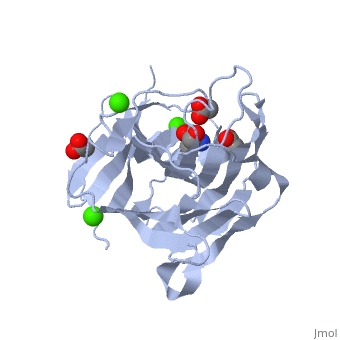
<StructureSection load='3h0o' size='340' side='right'caption='[[3h0o]], [[Resolution|resolution]] 1.40Å' scene=''> | <StructureSection load='3h0o' size='340' side='right'caption='[[3h0o]], [[Resolution|resolution]] 1.40Å' scene=''> | ||
== Structural highlights == | == Structural highlights == | ||
| - | <table><tr><td colspan='2'>[[3h0o]] is a 1 chain structure with sequence from [https://en.wikipedia.org/wiki/ | + | <table><tr><td colspan='2'>[[3h0o]] is a 1 chain structure with sequence from [https://en.wikipedia.org/wiki/Fibrobacter_succinogenes Fibrobacter succinogenes]. Full crystallographic information is available from [http://oca.weizmann.ac.il/oca-bin/ocashort?id=3H0O OCA]. For a <b>guided tour on the structure components</b> use [https://proteopedia.org/fgij/fg.htm?mol=3H0O FirstGlance]. <br> |
| - | </td></tr><tr id=' | + | </td></tr><tr id='method'><td class="sblockLbl"><b>[[Empirical_models|Method:]]</b></td><td class="sblockDat" id="methodDat">X-ray diffraction, [[Resolution|Resolution]] 1.4Å</td></tr> |
| - | <tr id=' | + | <tr id='ligand'><td class="sblockLbl"><b>[[Ligand|Ligands:]]</b></td><td class="sblockDat" id="ligandDat"><scene name='pdbligand=ACT:ACETATE+ION'>ACT</scene>, <scene name='pdbligand=CA:CALCIUM+ION'>CA</scene>, <scene name='pdbligand=TRS:2-AMINO-2-HYDROXYMETHYL-PROPANE-1,3-DIOL'>TRS</scene></td></tr> |
| - | + | ||
<tr id='resources'><td class="sblockLbl"><b>Resources:</b></td><td class="sblockDat"><span class='plainlinks'>[https://proteopedia.org/fgij/fg.htm?mol=3h0o FirstGlance], [http://oca.weizmann.ac.il/oca-bin/ocaids?id=3h0o OCA], [https://pdbe.org/3h0o PDBe], [https://www.rcsb.org/pdb/explore.do?structureId=3h0o RCSB], [https://www.ebi.ac.uk/pdbsum/3h0o PDBsum], [https://prosat.h-its.org/prosat/prosatexe?pdbcode=3h0o ProSAT]</span></td></tr> | <tr id='resources'><td class="sblockLbl"><b>Resources:</b></td><td class="sblockDat"><span class='plainlinks'>[https://proteopedia.org/fgij/fg.htm?mol=3h0o FirstGlance], [http://oca.weizmann.ac.il/oca-bin/ocaids?id=3h0o OCA], [https://pdbe.org/3h0o PDBe], [https://www.rcsb.org/pdb/explore.do?structureId=3h0o RCSB], [https://www.ebi.ac.uk/pdbsum/3h0o PDBsum], [https://prosat.h-its.org/prosat/prosatexe?pdbcode=3h0o ProSAT]</span></td></tr> | ||
</table> | </table> | ||
| + | == Function == | ||
| + | [https://www.uniprot.org/uniprot/GUB_FIBSS GUB_FIBSS] | ||
== Evolutionary Conservation == | == Evolutionary Conservation == | ||
[[Image:Consurf_key_small.gif|200px|right]] | [[Image:Consurf_key_small.gif|200px|right]] | ||
| Line 19: | Line 20: | ||
</jmol>, as determined by [http://consurfdb.tau.ac.il/ ConSurfDB]. You may read the [[Conservation%2C_Evolutionary|explanation]] of the method and the full data available from [http://bental.tau.ac.il/new_ConSurfDB/main_output.php?pdb_ID=3h0o ConSurf]. | </jmol>, as determined by [http://consurfdb.tau.ac.il/ ConSurfDB]. You may read the [[Conservation%2C_Evolutionary|explanation]] of the method and the full data available from [http://bental.tau.ac.il/new_ConSurfDB/main_output.php?pdb_ID=3h0o ConSurf]. | ||
<div style="clear:both"></div> | <div style="clear:both"></div> | ||
| - | <div style="background-color:#fffaf0;"> | ||
| - | == Publication Abstract from PubMed == | ||
| - | Fibrobacter succinogenes 1,3-1,4-beta-D-glucanase (Fsbeta-glucanase) catalyzes the specific hydrolysis of beta-1,4 glycosidic bonds adjacent to beta-1,3 linkages in beta-D-glucans or lichenan. This is the first report to elucidate the crystal structure of a truncated Fsbeta-glucanase (TFsbeta-glucanase) in complex with beta-1,3-1,4-cellotriose, a major product of the enzyme reaction. The crystal structures, at a resolution of 2.3 angstroms, reveal that the overall fold of TFsbeta-glucanase remains virtually unchanged upon sugar binding. The enzyme accommodates five glucose residues, forming a concave active cleft. The beta-1,3-1,4-cellotriose with subsites -3 to -1 bound to the active cleft of TFsbeta-glucanase with its reducing end subsite -1 close to the key catalytic residues Glu56 and Glu60. All three subsites of the beta-1,3-1,4-cellotriose adopted a relaxed C(1)4 conformation, with a beta-1,3 glycosidic linkage between subsites -2 and -1, and a beta-1,4 glycosidic linkage between subsites -3 and -2. On the basis of the enzyme-product complex structure observed in this study, a catalytic mechanism and substrate binding conformation of the active site of TFsbeta-glucanase is proposed. | ||
| - | |||
| - | Crystal structure of truncated Fibrobacter succinogenes 1,3-1,4-beta-D-glucanase in complex with beta-1,3-1,4-cellotriose.,Tsai LC, Shyur LF, Cheng YS, Lee SH J Mol Biol. 2005 Dec 2;354(3):642-51. Epub 2005 Sep 30. PMID:16246371<ref>PMID:16246371</ref> | ||
| - | |||
| - | From MEDLINE®/PubMed®, a database of the U.S. National Library of Medicine.<br> | ||
| - | </div> | ||
| - | <div class="pdbe-citations 3h0o" style="background-color:#fffaf0;"></div> | ||
==See Also== | ==See Also== | ||
*[[Glucanase 3D structures|Glucanase 3D structures]] | *[[Glucanase 3D structures|Glucanase 3D structures]] | ||
| - | == References == | ||
| - | <references/> | ||
__TOC__ | __TOC__ | ||
</StructureSection> | </StructureSection> | ||
| + | [[Category: Fibrobacter succinogenes]] | ||
[[Category: Large Structures]] | [[Category: Large Structures]] | ||
| - | + | [[Category: Hsiao CH]] | |
| - | [[Category: Hsiao | + | [[Category: Tsai LC]] |
| - | [[Category: Tsai | + | |
| - | + | ||
| - | + | ||
| - | + | ||
| - | + | ||
| - | + | ||
| - | + | ||
Current revision
The importance of CH-Pi stacking interactions between carbohydrate and aromatic residues in truncated Fibrobacter succinogenes 1,3-1,4-beta-D-glucanase
| |||||||||||