We apologize for Proteopedia being slow to respond. For the past two years, a new implementation of Proteopedia has been being built. Soon, it will replace this 18-year old system. All existing content will be moved to the new system at a date that will be announced here.
Sandbox Reserved 1725
From Proteopedia
(Difference between revisions)
| Line 16: | Line 16: | ||
===Overview=== | ===Overview=== | ||
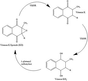
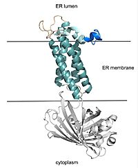
| - | + | There are 4 catalytic cysteines that are important to VKOR, 43, 51, 132, and 135. To explain how this works, it is easiest to start with the second state. In the second state, an oxidized or partially oxidized Vitamin K has entered the active site. The stabilizing 51-132 disulfide bond is shown. Then in the third state, 43 has attacked the disulfide bond and made its own bond with 51. You can see 132 has an oxygen. That is because the researchers made a mutation from S to O to force the reaction to stop at that step so the structure could be deduced. In the natural VKOR, that would be a sulfur. The next state, the open state, results from 132 forming a bridge with 135. This allows release of the reduced or partially reduced Vitamin K. All of this disulfide rearranging was working to reduce the Vitamin K, particularly in the 135 position. If we go back to State 2, when Vitamin K first binds, you can see that 135 is not tied up in a disulfide bond. It is available to help the Vitamin K bond. So, it makes sense that once 135 gets forced to bond, the now reduced Vitamin K is released. State 5 is interesting because the disulfide bonds are similar to the open state, but warfarin is actually bound. This represents the binding of warfarin to the fully oxidized VKOR at the end of its cycle. Going back to State 1, the researchers used a mutation at 43 to mimic VKOR’s partially oxidized state. Warfarin can also bind to this state and notice that the disulfide bonds are the same as State 2. Also it is worth pointing out how the disulfide bonds contribute to conformational changes and are affected by conformational changes, which affects their proximity to each other and the active site. | |
| + | |||
===Catalytic Cysteines=== | ===Catalytic Cysteines=== | ||
| - | |||
VKOR uses four catalytic cysteines (43, 51, 132, and 135) to facilitate reduction and cause conformational changes via disulfide bridge formation. When an oxidized or partially oxidized Vitamin K enters the active site, VKOR has a stabilizing C51-C132 disulfide bond, known as the closed state. C135 is not in a disulfide bridge and is important in helping Vitamin K bind. C43 attacks the C51-C132 bond, forming a new C43-C51 bond that characterizes the open state. The free amino acid at the 132 position is shown as a serine in this depiction because it was mutated it to stop the reaction at this intermediate during Liu's experimental procedure. <ref name="Liu">PMID:33154105</ref>. In the wild type VKOR, the 132 position would be a cysteine. C132 forms a bridge with C135 which allows release of the reduced or partially reduced Vitamin K. These rearrangements facilitate the use of cysteines as reducing agents for Vitamin K. Cysteine rearrangements also contribute to and are affected by overall conformational changes which affects their proximity to each other and the active site. | VKOR uses four catalytic cysteines (43, 51, 132, and 135) to facilitate reduction and cause conformational changes via disulfide bridge formation. When an oxidized or partially oxidized Vitamin K enters the active site, VKOR has a stabilizing C51-C132 disulfide bond, known as the closed state. C135 is not in a disulfide bridge and is important in helping Vitamin K bind. C43 attacks the C51-C132 bond, forming a new C43-C51 bond that characterizes the open state. The free amino acid at the 132 position is shown as a serine in this depiction because it was mutated it to stop the reaction at this intermediate during Liu's experimental procedure. <ref name="Liu">PMID:33154105</ref>. In the wild type VKOR, the 132 position would be a cysteine. C132 forms a bridge with C135 which allows release of the reduced or partially reduced Vitamin K. These rearrangements facilitate the use of cysteines as reducing agents for Vitamin K. Cysteine rearrangements also contribute to and are affected by overall conformational changes which affects their proximity to each other and the active site. | ||
Warfarin binding also depends on the catalytic cysteines. Warfarin is able to bind to the fully oxidized form of VKOR where the disulfide bridge pairings are C132-C135 and C43-C51. Warfarin can also bind to the partially oxidized form of VKOR where the disulfide bridge pairings are the same as the closed state, with C51-C132 being the only bridge. Again, C43 is shown as a serine because a mutation was used to force VKOR to adopt that conformation during the experimental procedure by Liu. <ref name="Liu">PMID:33154105</ref>. | Warfarin binding also depends on the catalytic cysteines. Warfarin is able to bind to the fully oxidized form of VKOR where the disulfide bridge pairings are C132-C135 and C43-C51. Warfarin can also bind to the partially oxidized form of VKOR where the disulfide bridge pairings are the same as the closed state, with C51-C132 being the only bridge. Again, C43 is shown as a serine because a mutation was used to force VKOR to adopt that conformation during the experimental procedure by Liu. <ref name="Liu">PMID:33154105</ref>. | ||
| - | |||
=== Catalytic Amino Acids === | === Catalytic Amino Acids === | ||
| + | VKOR uses two catalytic amino acids, tyrosine 139 and asparagine 80, to stabilize <scene name='90/904329/Kohhbond/1'>vitamin K</scene> in all forms and <scene name='90/904329/Warfarinhbond/2'>vitamin K antagonists</scene>, such as warfarin, in the binding pocket. Tyr139 and Asn80 hydrogen bond to carbonyl groups on both structures and stabilizes them within the binding pocket. | ||
| + | === Hydrophobic Interactions === | ||
| - | |||
| - | === Hydrophobic Interactions === | ||
== Medical Relevance == | == Medical Relevance == | ||
Revision as of 20:54, 24 March 2022
| This Sandbox is Reserved from February 28 through September 1, 2022 for use in the course CH462 Biochemistry II taught by R. Jeremy Johnson at the Butler University, Indianapolis, USA. This reservation includes Sandbox Reserved 1700 through Sandbox Reserved 1729. |
To get started:
More help: Help:Editing |
Vitamin K Epoxide Reductase
| |||||||||||
References
- ↑ Stafford DW. The vitamin K cycle. J Thromb Haemost. 2005 Aug;3(8):1873-8. doi: 10.1111/j.1538-7836.2005.01419.x. PMID:16102054 doi:http://dx.doi.org/10.1111/j.1538-7836.2005.01419.x
- ↑ 2.0 2.1 2.2 Liu S, Li S, Shen G, Sukumar N, Krezel AM, Li W. Structural basis of antagonizing the vitamin K catalytic cycle for anticoagulation. Science. 2020 Nov 5. pii: science.abc5667. doi: 10.1126/science.abc5667. PMID:33154105 doi:http://dx.doi.org/10.1126/science.abc5667
Student Contributors
Izabella Jordan, Emma Varness


