We apologize for Proteopedia being slow to respond. For the past two years, a new implementation of Proteopedia has been being built. Soon, it will replace this 18-year old system. All existing content will be moved to the new system at a date that will be announced here.
Sandbox Reserved 1709
From Proteopedia
(Difference between revisions)
| Line 26: | Line 26: | ||
=== Anchor === | === Anchor === | ||
<scene name='90/904314/Anchor_domain/1'>Anchor Domain</scene> | <scene name='90/904314/Anchor_domain/1'>Anchor Domain</scene> | ||
| + | |||
== Function: Method of Coagulation == | == Function: Method of Coagulation == | ||
| + | |||
=== Brief Overview === | === Brief Overview === | ||
The <scene name='90/906893/Open_conformation/1'>open conformation</scene> will be prepped and waiting for a substrate or ligand to bind. | The <scene name='90/906893/Open_conformation/1'>open conformation</scene> will be prepped and waiting for a substrate or ligand to bind. | ||
| + | |||
=== Catalytic Mechanism === | === Catalytic Mechanism === | ||
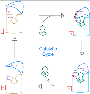
[[Image:Catalytic Mech Pic.png |350 px| right| thumb]] | [[Image:Catalytic Mech Pic.png |350 px| right| thumb]] | ||
| Line 34: | Line 37: | ||
The catalytic mechanism of VKOR is a critical part of its overall function in the body. Highly regulated enzymatic activity through the reactivity of catalytic cysteines allows VKOR to properly activate Vitamin K for its use in the body. The enzyme begins in <scene name='90/906893/Stage_1_catalytic_cycle/1'>stage 1</scene>, where it's in the open conformation with the cap domain open to allow in a substrate to bind to the active site. Once a substrate binds, the cap domain is initiated into the closed conformation. VKOR is now in <scene name='90/906893/Stage_2_catalytic_cycle/1'>stage 2</scene>. To further stabilize the closed conformation with the substrate bound, the cap domain helps initiate a catalytic reaction of cysteines to break the disulfide bridge that was stabilizing stage 1. Free cysteines are now available that provide strong stabilization of the closed conformation through interactions with the cap domain and the bound substrate. This puts the enzyme in <scene name='90/904314/Stage_3_catalytic_cycle/1'>Stage 3</scene>, where the catalytic free cysteines react to form a new disulfide bridge, releasing the activated substrate into the blood stream to promote anticoagulation. With two stable disulfide bridges and VKOR unbound, the enzyme is now in its final, unreactive <scene name='90/904314/Stage_4_catalytic_cycle/1'>Stage 4</scene>. VKOR must undergo conformational changes to return to Stage 1 and restart the catalytic process to activate Vitamin K again. | The catalytic mechanism of VKOR is a critical part of its overall function in the body. Highly regulated enzymatic activity through the reactivity of catalytic cysteines allows VKOR to properly activate Vitamin K for its use in the body. The enzyme begins in <scene name='90/906893/Stage_1_catalytic_cycle/1'>stage 1</scene>, where it's in the open conformation with the cap domain open to allow in a substrate to bind to the active site. Once a substrate binds, the cap domain is initiated into the closed conformation. VKOR is now in <scene name='90/906893/Stage_2_catalytic_cycle/1'>stage 2</scene>. To further stabilize the closed conformation with the substrate bound, the cap domain helps initiate a catalytic reaction of cysteines to break the disulfide bridge that was stabilizing stage 1. Free cysteines are now available that provide strong stabilization of the closed conformation through interactions with the cap domain and the bound substrate. This puts the enzyme in <scene name='90/904314/Stage_3_catalytic_cycle/1'>Stage 3</scene>, where the catalytic free cysteines react to form a new disulfide bridge, releasing the activated substrate into the blood stream to promote anticoagulation. With two stable disulfide bridges and VKOR unbound, the enzyme is now in its final, unreactive <scene name='90/904314/Stage_4_catalytic_cycle/1'>Stage 4</scene>. VKOR must undergo conformational changes to return to Stage 1 and restart the catalytic process to activate Vitamin K again. | ||
[[Image:VKOR_Stage_3_and_4_mechanism.png |400 px|right| thumb]] | [[Image:VKOR_Stage_3_and_4_mechanism.png |400 px|right| thumb]] | ||
| + | |||
== Disease and Treatment == | == Disease and Treatment == | ||
=== Afflictions === | === Afflictions === | ||
=== Inhibition === | === Inhibition === | ||
| - | The most inexpensive and common way to treat blood clotting is through the VKOR inhibitor, <scene name='90/906893/Vkor_with_warfarin_bound/1'>Warfarin</scene>. | + | The most inexpensive and common way to treat blood clotting is through the VKOR inhibitor, <scene name='90/906893/Vkor_with_warfarin_bound/1'>Warfarin</scene>. [https://en.wikipedia.org/wiki/Warfarin Warfarin] is able to do so by outcompeting KO. It will enter the binding pocket of VKOR, creating strong hydrogen bonds with the active site. |
| + | |||
=== Mutations === | === Mutations === | ||
Some key <scene name='90/906893/Active_site_mutations/1'>mutations</scene> that can be detrimental to the VKOR structure are mutations of the <scene name='90/906893/Active_site/2'>active site</scene>. The two main residues, N80 and Y139, can be mutated to A80 and F139 creating a decrease in recognition and stabilization | Some key <scene name='90/906893/Active_site_mutations/1'>mutations</scene> that can be detrimental to the VKOR structure are mutations of the <scene name='90/906893/Active_site/2'>active site</scene>. The two main residues, N80 and Y139, can be mutated to A80 and F139 creating a decrease in recognition and stabilization | ||
| Line 44: | Line 49: | ||
</StructureSection> | </StructureSection> | ||
| + | |||
== References == | == References == | ||
1. Li, Weikai et al. “Structure of a bacterial homologue of vitamin K epoxide reductase.” Nature vol. 463,7280 (2010): 507-12. [https://www.ncbi.nlm.nih.gov/pmc/articles/PMC2919313/ doi:10.1038/nature08720]. | 1. Li, Weikai et al. “Structure of a bacterial homologue of vitamin K epoxide reductase.” Nature vol. 463,7280 (2010): 507-12. [https://www.ncbi.nlm.nih.gov/pmc/articles/PMC2919313/ doi:10.1038/nature08720]. | ||
Revision as of 22:18, 28 March 2022
| |||||||||||
References
1. Li, Weikai et al. “Structure of a bacterial homologue of vitamin K epoxide reductase.” Nature vol. 463,7280 (2010): 507-12. doi:10.1038/nature08720.
2. Liu S, Li S, Shen G, Sukumar N, Krezel AM, Li W. Structural basis of antagonizing the vitamin K catalytic cycle for anticoagulation. Science. 2021 Jan 1;371(6524):eabc5667. doi: 10.1126/science.abc5667. Epub 2020 Nov 5. PMID: 33154105; PMCID: PMC7946407.
3. “Warfarin.” Wikipedia, Wikimedia Foundation, 10 Feb. 2022, https://en.wikipedia.org/wiki/Warfarin.
- ↑ Hanson, R. M., Prilusky, J., Renjian, Z., Nakane, T. and Sussman, J. L. (2013), JSmol and the Next-Generation Web-Based Representation of 3D Molecular Structure as Applied to Proteopedia. Isr. J. Chem., 53:207-216. doi:http://dx.doi.org/10.1002/ijch.201300024
- ↑ Herraez A. Biomolecules in the computer: Jmol to the rescue. Biochem Mol Biol Educ. 2006 Jul;34(4):255-61. doi: 10.1002/bmb.2006.494034042644. PMID:21638687 doi:10.1002/bmb.2006.494034042644


